Table of Contents
Advertisement
English
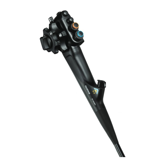
Video Endoscope
EI-580BT
OPERATION MANUAL
This Operation Manual describes details on how to operate the video endoscope and cautions to be
observed when operating it. Please read this manual thoroughly before actually operating the video
endoscope.
After reading this manual, store it nearby the video endoscope so that you can see it whenever
necessary.
202B1263075
Advertisement
Table of Contents

Related Manuals for FujiFilm EI-580BT


Summary of Contents for FujiFilm EI-580BT
- Page 1 English Video Endoscope EI-580BT OPERATION MANUAL This Operation Manual describes details on how to operate the video endoscope and cautions to be observed when operating it. Please read this manual thoroughly before actually operating the video endoscope. After reading this manual, store it nearby the video endoscope so that you can see it whenever necessary.
- Page 2 202B1263075...
-
Page 3: Introduction
Introduction Introduction Video Endoscope EI-580BT is a medical endoscope intended for the observation, diagnosis and endoscopic treatment of the esophagus, stomach, duodenum, small intestine, ileocecal region, large intestine, sigmoid colon and rectum at medical facilities under the management of physicians. - Page 4 FUJIFILM Corporation. FUJIFILM Corporation shall not be liable for malfunctions and damages of FUJIFILM Corporation products due to products of other manufacturers not supplied by FUJIFILM Corporation.
-
Page 5: Endoscope Manuals
This manual describes the cleaning, disinfection, sterilization and storage methods of the video endoscope. [Note] In this manual, the Video Endoscope EI-580BT Operation Manual is referred to as “this manual”, and the Video Endoscope EI-580BT Reprocessing Manual as “Reprocessing Manual”. -
Page 6: Contents At A Glance
Contents at a Glance Contents at a Glance Chapter 1 Precautions This chapter describes the warnings and cautions for safe operation of the endoscope. Chapter 2 Product Overview This chapter describes the composition of the endoscope set of accessories and system configuration. Chapter 3 Preparation and Inspection Before Use This chapter describes the inspection and preparation methods to be... -
Page 7: Table Of Contents
Contents Contents Introduction ........................3 Endoscope Manuals .....................5 How to Read This Manual ...................5 Contents at a Glance ....................6 Chapter 1 Precautions ....................10 1.1 For Safe Operation ...............10 1.2 Classification ................10 1.3 Safety ....................11 1.3.1 Infection ................11 1.3.2 Preventing Electrical Shock ..........14 1.3.3 Treatment with Electrosurgical Instruments ....14 1.3.4... - Page 8 Contents 2.6 Operating Bending Section ............40 2.6.1 Operating the Bending Section ........40 2.6.2 Angle Lock Function ............42 2.7 Control Valves ................44 2.7.1 Suction Valve ..............44 2.7.2 Air/Water Valve ..............44 2.8 Scope Switches for Images and Recording ........45 Chapter 3 Preparation and Inspection Before Use ............46 3.1 Preparing Forceps Valve ..............46 3.1.1 Cleaning and Disinfecting (or Sterilizing)
- Page 9 Contents Chapter 4 Method of Use ..................74 4.1 Preparation..................76 4.1.1 Preparing Necessary Equipment ........76 4.1.2 Pretreatment of the Patient ..........76 4.2 Insertion and Observation ............77 4.2.1 Peroral Insertion ..............79 4.2.2 Transanal Insertion ............83 4.2.3 To Suck Mucus ...............87 4.2.4 If Mucus Adheres to the Distal Objective Lens or If the Image is Obscured ..........87 4.3 Treatment..................88 4.4 Endoscope Withdrawal ..............89...
-
Page 10: Chapter 1 Precautions
Do not modify this product or its components, and do not disassemble, repair or in any other way reverse-engineer these products. Even if you find a defect, do not attempt to repair these products yourself. FUJIFILM Corporation shall not be liable for any defects or device failures caused by such modifications, disassembly, repairs or reverse-engineering. -
Page 11: Safety
Chapter 1 Precautions 1.3 Safety Read the following precautions before using this product to ensure proper handling. 1.3.1 Infection WARNING This product has not been cleaned or disinfected (or sterilized). It must be cleaned and disinfected (or sterilized) for the first time prior to use and after any subsequent use as per instructions provided in the Reprocessing Manual. - Page 12 Chapter 1 Precautions WARNING The forceps valve must be completely immersed in a disinfectant solution. Remove air bubbles completely. If any air bubbles remain, effective disinfection cannot be achieved and an inadequately cleaned and disinfected (or sterilized) forceps valve may increase a risk of infection.
- Page 13 Chapter 1 Precautions WARNING Slowly insert an endotherapy device (e.g. forceps) or syringe straight into the endoscope. Also, when withdrawing it, slowly pull straight out. If it is inserted or withdrawn quickly, body fluid may be splattered around due to breakage or accidental detachment of the instrument, leading to infection.
-
Page 14: Preventing Electrical Shock
Chapter 1 Precautions 1.3.2 Preventing Electrical Shock WARNING Insert the AC plug into a hospital grade receptacle. Not doing so may cause an electric shock accident. Use an electrosurgical instrument conforming to EN 60601-2-2. Refer to the manual of the electrosurgical instrument for how to operate the electrosurgical instrument. - Page 15 Chapter 1 Precautions WARNING Wear electrically insulating gloves when using an electrosurgical instrument or accessory. If not worn, there is a risk of thermal injury or electric shock. Always keep pacemaker users away from electrosurgical instruments. The operation of the pacemaker will be malfunctioned by the electrosurgical instruments.
-
Page 16: Direct Harm To Human Body
Chapter 1 Precautions 1.3.4 Direct Harm to Human Body WARNING Use the air/water channel cleaning adapter CA-611 only for cleaning the air/water channel. If it is used during an examination or treatment, continuous air supply may occur and cause patient injury. Do not supply an excessive amount of air or gas during the procedure as doing so could cause an embolism. -
Page 17: Balloon Instructions
Chapter 1 Precautions CAUTION Set the suction pressure between 40 and 53 kPa. If the suction pressure is too high, the endoscope may adhere to mucous membrane, resulting in damage to the mucous membrane. Do not look directly into the light coming from the light guide at the distal end of the endoscope. - Page 18 Chapter 1 Precautions WARNING Insert and withdraw the endoscope or Over-tube slowly. Slowly insert the Over-tube so as not to have the intestinal tract caught in a gap between the endoscope and Over-tube. When holding the Over- tube, take care not to fold or crush the air feed inlet (clear tube). Do not press the endoscope or Over-tube strongly onto the walls of the digestive tract.
-
Page 19: Electromagnetic Compatibility (Emc)
• Consult the manufacturer or dealer of other devices. If the problem cannot be solved with the above measures, stop using this product and consult the manufacturer or your local FUJIFILM dealer for help. WARNING Do not place any objects that emit strong electromagnetic waves near this product. -
Page 20: Location Of Each Label
Chapter 1 Precautions 1.5 Location of Each Label The positions where the labels are affixed on this product are shown below. The relevant safety signs are also described. 1.5.1 Location of Labels <Control Portion> Model Label Instrument Channel Diameter Label <LG Connector>... -
Page 21: Symbols
Chapter 1 Precautions 1.5.2 Symbols Symbol Location Description Individual packaging Do not re-use / bag for accessories Single patient use only Individual packaging Use by date / Expiration date of use bag for accessories Individual packaging Lot number bag for accessories LG connector Serial number LG connector... -
Page 22: Possible Combinations For Use
Chapter 1 Precautions 1.6 Possible Combinations for Use 1.6.1 Accessories Use this product in combination with the accessories described in “Main Specification”. WARNING Do not use the endoscope in combination with accessories other than those described in this manual. Otherwise, it is unable to ensure its functionality, and creates a risk of patient injury or equipment damage. -
Page 23: Peripheral Devices
Read the operation manuals of the peripheral devices used in combination with this product. CAUTION For endoscopic CO regulators, use only those designated by FUJIFILM. If other devices are used, the air/water supply function may deteriorate, and it may not be possible to clean the lens properly. 202B1263075... -
Page 24: Cautions/Warnings
If any abnormality is noticed during use, carry out safety checks and discontinue use immediately. If any abnormality occurs with this product, refer to “Chapter 5 Troubleshooting”. Should any questions arise regarding information contained in the operation manuals or any safety concerns, contact your local FUJIFILM dealer. 202B1263075... -
Page 25: Transportation And Storage
Chapter 1 Precautions 1.7.2 Transportation and Storage WARNING Contact your local FUJIFILM dealer when this product is returned for repair. Be sure to clean and disinfect (or sterilize) this product before returning for repair. A returned product which is not cleaned and disinfected (or sterilized) may increase infection control risks to users, service personnel or other persons in contact with it. -
Page 26: General Warnings
Chapter 1 Precautions 1.7.4 General Warnings WARNING Make sure to inspect the equipment before use according to the procedures provided in this manual to avoid unexpected accidents and to take full advantage of the equipment’s capabilities. If the inspection result shows any abnormality, do not use the same equipment. This product is a medical endoscope intended for the observation, diagnosis and endoscopic treatment of the esophagus, stomach, duodenum, small intestine, rectum, sigmoid colon, large intestine and... - Page 27 Chapter 1 Precautions 202B1263075...
-
Page 28: Chapter 2 Product Overview
Chapter 2 Product Overview Chapter 2 Product Overview 2.1 Composition of Standard Set The endoscope standard set is provided in the carrying case and consists of the following items. [Note] Figures in parentheses indicate the number of articles. Carrying Case (1) Manuals Operation Manual (1) Reprocessing Manual (1) - Page 29 Chapter 2 Product Overview Air/water channel cleaning adapter Balloon Channel Cleaning Adapter CA-611 (1) CA-606 (1) Injection Tube Channel Plug WA-007 WA-010 Cleaning Adapter CA-610 (1) Cleaning Brush Cleaning Brush Ventilation Adapter WB7024FW (1) WB11003FW (1) AD-7 (1) 202B1263075...
-
Page 30: System Configuration
• Recording video images • Printing still images Water Tank WT-2 WT-4 Sonoprobe System Endoscopic CO Light Source Regulator XL-4450 GW-1 GW-100 Endoscope EI-580BT Video Processor VP-4450HD Balloon Foot Switch BS-2 BS-4 Data Keyboard DK-4450E Balloon Controller Over-tube PB-20 TS-13101... - Page 31 Chapter 2 Product Overview [Note] The devices described here may have already been discontinued. For details on the devices used in combination with this product, contact your local FUJIFILM dealer. [Note 1] Electrosurgical Unit ICC 200 (ERBE) [Note 1] For details, refer to the manual of the electrosurgical instrument.
-
Page 32: Combination With Vp-7000
• Printing still images Water Tank WT-2 WT-4 Sonoprobe System Endoscopic CO Regulator Light Source GW-1 [Note 1] BL-7000 GW-100 Endoscope EI-580BT Video Processor [Note 1] VP-7000 Balloon Controller Balloon PB-20 BS-2 PB-30 BS-4 Data Keyboard DK-7000U Over-tube [Note 2]... - Page 33 [Note] The devices described here may have already been discontinued. For details on the devices used in combination with this product, contact your local FUJIFILM dealer. [Note 1] For this endoscope, place the VP-7000 video processor on the lower shelf and the BL-7000 light source on the upper shelf.
-
Page 34: Nomenclature And Functions Of Endoscope
Chapter 2 Product Overview 2.3 Nomenclature and Functions of Endoscope This product consists of the following parts. [Note] Use the video processor to assign functions on each scope switch. For details, see the operation manual of the video processor. LG Flexible Portion Contains light guide, air/water supply tube, suction tube and cables. - Page 35 Chapter 2 Product Overview [Note 1] Scope Switch 2 [Note 1] Scope Switch 1 Up-down Angulation Lock Suction Valve Locks the up/down angulation of the bending section. Allows suction through instrument channel (port) from the distal end. This Up-down Angulation Knob valve has an orange mark.
-
Page 36: Nomenclature And Functions Of Endoscope Distal End
Chapter 2 Product Overview 2.4 Nomenclature and Functions of Endoscope Distal End Imaging Section (Inside) Cover Light Guide The light from the light source is emitted Balloon Air Feed Outlet from these windows. The air supplied from the balloon air feed inlet exits from this outlet. -
Page 37: Nomenclature And Functions Of Accessories
Chapter 2 Product Overview 2.5 Nomenclature and Functions of Accessories 2.5.1 Forceps Valve The forceps valve is provided without sterilization and must be cleaned and disinfected (or sterilized) prior to use following the instructions of the Reprocessing Manual. Reprocessing Manual “7.1 Cleaning and Disinfecting (or Sterilizing) Forceps Valve” Slit Circular Hole Entire Forceps Valve... -
Page 38: Cleaning Adapter
Chapter 2 Product Overview 2.5.2 Cleaning Adapter The cleaning adapter (CA-610) is comprised of the injection tube (WA-007) and channel plug (WA-010). <Injection Tube (WA-007)> Tube for Instrument/Suction Channel Connects to the suction channel. Air Supply Tube Port Connects to the air supply Air/Water Channel Connector tube of the LG connector on the endoscope. - Page 39 Chapter 2 Product Overview <Channel Plug (WA-010)> Suction Plug This plug is inserted into the Plug Frame suction valve cylinder of the Connects to the valve on the endoscope. endoscope's control portion. Air/Water Plug This plug is inserted into the air/water valve cylinder of the endoscope.
-
Page 40: Operating Bending Section
Chapter 2 Product Overview 2.6 Operating Bending Section 2.6.1 Operating the Bending Section CAUTION Do not forcibly twist or bend too sharply the insertion tube and the bending section by hand. It may cause a failure. The bending section is operated by using up-down/left-right angulation knobs. <Up-down Angulation Knob>... - Page 41 Chapter 2 Product Overview <Left-right Angulation Knob> Turn the left-right angulation knob in the direction of L to angulate the bending section to the left. Turn it in the direction of R to angulate the bending section to the right. 202B1263075...
-
Page 42: Angle Lock Function
Chapter 2 Product Overview 2.6.2 Angle Lock Function This angle lock function is used to maintain a bent condition of the bending section. With the up-down angulation lock operation, it switches between lock and free (unlock) of the up/down angulation. With the left-right angulation lock operation, it switches between lock and free (unlock) of the left/right angulation. - Page 43 Chapter 2 Product Overview <Left-right Angulation Lock> Rotate the knob in the direction of F to unlock the bending section. [Note] Free (unlock) : Allows external force to angulate the bending section freely. Rotate the knob in the direction opposite to F to lock the bending section.
-
Page 44: Control Valves
Chapter 2 Product Overview 2.7 Control Valves 2.7.1 Suction Valve Suction is activated while this valve is depressed. Suction through the instrument channel requires the forceps valve be attached to the instrument channel inlet. 2.7.2 Air/Water Valve To supply air, cover the hole in the center of this valve with one’s finger. -
Page 45: Scope Switches For Images And Recording
Chapter 2 Product Overview 2.8 Scope Switches for Images and Recording CAUTION Use the video processor to assign functions on each scope switch. For details, see the operation manual of the video processor. When one switch is pressed, the function assigned to it activates. Scope Switches 4 Scope Switches 3 Scope Switches 2... -
Page 46: Chapter 3 Preparation And Inspection Before Use
Chapter 3 Preparation and Inspection Before Use Chapter 3 Preparation and Inspection Before Use WARNING Make sure to inspect the equipment before use according to the procedures provided in this manual to avoid unexpected accidents and to take full advantage of the equipment’s capabilities. If the inspection result shows any abnormality, do not use the same equipment. -
Page 47: Cleaning And Disinfecting (Or Sterilizing) The Forceps Valve
Chapter 3 Preparation and Inspection Before Use 3.1.1 Cleaning and Disinfecting (or Sterilizing) the Forceps Valve The forceps valve is provided without sterilization and must be cleaned and disinfected (or sterilized) prior to use following the instructions of the Reprocessing Manual. The forceps valve is a single-use product. -
Page 48: Preparing Air/Water Valve And Suction Valve
Chapter 3 Preparation and Inspection Before Use 3.2 Preparing Air/Water Valve and Suction Valve WARNING Use a cleaned and disinfected (or sterilized) air/water valve and suction valve. An inadequately cleaned and disinfected (or sterilized) valve may pose an infection risk. Prepare an appropriately cleaned and disinfected (or sterilized) air/water valve and suction valve. -
Page 49: Attaching The Air/Water Valve And Suction Valve
Chapter 3 Preparation and Inspection Before Use 3.2.2 Attaching the Air/Water Valve and Suction Valve Attach the air/water valve and suction valve to the control portion of the endoscope. (1) Attach the air/water valve to the endoscope’s air/water valve cylinder and push in the valve firmly. [Note] The air/water valve and the air/water valve cylinder have a blue mark. -
Page 50: Preparing And Inspecting Over-Tube And Balloon
Chapter 3 Preparation and Inspection Before Use 3.3 Preparing and Inspecting Over-tube and Balloon WARNING Use a sterilized balloon installation tool. Do not use the balloon or Over-tube after its expiration date has elapsed. Doing so may pose an infection risk. Do not use the TS-13101 Over-tubes and BS-2 balloons on patients allergic to latex. - Page 51 Chapter 3 Preparation and Inspection Before Use (2) Prepare the fixing rubber setting tool and balloon setting tool for the balloon to be used. Table 3.3 Compatible setting tools with the balloons Fixing rubber Balloon Balloon setting tool setting tool ST-01B BS-2 ST-05B...
-
Page 52: Attaching The Over-Tube
Chapter 3 Preparation and Inspection Before Use 3.3.2 Attaching the Over-tube (1) Moisten the inside of Over-tube by injecting sterile water into the water feed inlet (blue tube) using a syringe. Check valve (2) Insert the endoscope from the endoscope insertion inlet of Cover rubber the Over-tube to the proximal end of the flexible portion. - Page 53 Chapter 3 Preparation and Inspection Before Use (2) Check that the tip of the endoscope is free of dirt. Apply a small amount of 70% ethanol solution to the tip of the endoscope. (3) Apply a small amount of 70% ethanol solution to the outside of the balloon setting tool (ST-05B).
-
Page 54: Attaching The Fixing Rubber
Chapter 3 Preparation and Inspection Before Use (6) Roll up the edge of the balloon, and slide the balloon setting tool (ST-05B) toward the control portion of the endoscope to mount the balloon on the endoscope. Return the edge of the balloon to the original form. [Note] Make the ethanol inside the balloon evaporate by widening the end of the balloon. - Page 55 Chapter 3 Preparation and Inspection Before Use (2) Slide the fixing rubber down from the fixing rubber setting tool (ST-01B) to the end of the balloon on the curved section side. (3) When using a hood, attach it according to “3.3.4 Attaching the Hood”.
- Page 56 Chapter 3 Preparation and Inspection Before Use (3) Attach a fixing rubber to the tip of the closed claw section. [Note] Deeply insert the claw section into a fixing rubber until the tip of the claw section protrudes from the fixing rubber. (4) Slowly pull the trigger until the end surface of the piston makes contact with the fixing rubber.
-
Page 57: Attaching The Hood (Only When Necessary)
Chapter 3 Preparation and Inspection Before Use (7) While opening the claw section by pulling the trigger, withdraw the setting tool from the distal end of endoscope. (8) When using a hood, attach it according to “3.3.4 Attaching the Hood”. “3.3.4 Attaching the Hood”... - Page 58 Chapter 3 Preparation and Inspection Before Use (1) Attach the balloon according to “3.3.3 Mounting the Endoscope Balloon.” (2) Attach a hood (DH-17EN) onto the distal end of endoscope. Insert the endoscope until it reaches the end of the hood. [Note] Attach a hood onto the dry distal end of endoscope.
-
Page 59: Inspecting The Balloon
Chapter 3 Preparation and Inspection Before Use 3.3.6 Inspecting the Balloon (1) Supply air into the balloon air feed inlet to make sure that the balloon attached to the endoscope inflates. (2) Remove air from the balloon air feed inlet to make sure that the balloon deflates. -
Page 60: Connecting Balloon Controller
Chapter 3 Preparation and Inspection Before Use 3.4 Connecting Balloon Controller WARNING The filter and tube of the tube kit are consumables. Replace them once a month or once every 10 cases, whichever comes first. If body fluid flows back into the tube, replace the tube kit. Not doing so may pose an infection risk. -
Page 61: Inspecting The Balloon (Balloon Controller)
Chapter 3 Preparation and Inspection Before Use (2) Securely insert the endoscope-side connector of the tube kit (TY-06S) into the balloon air feed inlet of the endoscope. (3) Connect the tube 1 (white tube) of the balloon controller to the air feed inlet (clear tube) of the Over-tube. 3.4.2 Inspecting the Balloon (Balloon Controller) (1) Make sure that the “ENDOSCOPE”... - Page 62 Chapter 3 Preparation and Inspection Before Use (4) Press the Over-tube-side switch on the balloon controller to make sure that the balloon attached to the distal end of Over-tube inflates. (5) Press the switch again to make sure that the balloon deflates.
-
Page 63: Preparing System
Chapter 3 Preparation and Inspection Before Use 3.5 Preparing System WARNING When supplying water, use sterile water. If sterile water is not used, it can create a risk of infection. 3.5.1 Preparing Peripheral Devices For details on preparation and inspection of peripheral devices, refer to their manuals. 3.5.2 Preparing the System (1) Move the cart with the video processor, light source, balloon controller and other peripherals to the place where... - Page 64 Chapter 3 Preparation and Inspection Before Use (3) Prepare the suction unit. [Note] For details on the suction unit, refer to the manual of the suction unit. Collection canister Suction unit (4) Mount the water tank, 80% filled with sterile water, on the hook.
-
Page 65: Connecting Endoscope (Attachment)
Chapter 3 Preparation and Inspection Before Use 3.6 Connecting Endoscope (Attachment) WARNING Firmly connect the scope connector of the endoscope and the light source. If the scope connector is not connected properly, the endoscopic image may flicker or be lost. CAUTION Do not attach the video connector of the endoscope to the electrical connector socket of the VP-7000 with the endoscope inserted into the... - Page 66 Chapter 3 Preparation and Inspection Before Use (2) Insert the LG connector of the endoscope into the endoscope socket on the light source until it stops. (3) Insert the video connector of the endoscope into the video connector socket. Align the mark of the connector with the one of the connector socket, and attach the connector firmly by turning it clockwise while slightly pushing it.
-
Page 67: Connecting The Water Tank And Suction Unit
Chapter 3 Preparation and Inspection Before Use In alignment with the video connector index, rotate the connector clockwise while lightly pressing it. [Note] Make sure to rotate the video connector index 90 degrees clockwise. [Note] Do not connect more than one endoscope. 3.6.2 Connecting the Water Tank and Suction Unit (1) Make sure that the forceps valve is attached to the instrument channel inlet the endoscope. -
Page 68: Inspecting Endoscope
If the adhesive surfaces are rough, pitted or flaking, return the instrument to your local FUJIFILM dealer. (2) Hold the insertion tube with both hands to make a semicircle with a diameter of approximately 200 mm, referring to the figure at right. -
Page 69: Inspecting The Bending Mechanism
Chapter 3 Preparation and Inspection Before Use 3.7.2 Inspecting the Bending Mechanism [Note] For information on how to operate the bending mechanism, refer to “2.6 Operating Bending Section” of this manual. “2.6 Operating Bending Section” (1) Unlock both up-down and left-right angulation locks by turning them in the “Free”... - Page 70 Chapter 3 Preparation and Inspection Before Use (3) Place the distal end of the endoscope without touching the floor, etc., depress the air/water valve, and check that sterile water comes out of the nozzle. [Note] Note that the direction in which sterile water comes out. [Note] Manipulation method differs when a gas/water button provided with an endoscopic CO...
-
Page 71: Inspecting The Balloon Air Feed Inlet
Chapter 3 Preparation and Inspection Before Use 3.7.4 Inspecting the Balloon Air Feed Inlet Supply air from the balloon air feed inlet with a syringe and make sure that the air exits from the balloon air feed outlet. 3.7.5 Inspecting the Instrument Channel Insert an endotherapy device from the instrument channel inlet and check that the tip of endotherapy device comes smoothly out of the instrument channel outlet in the distal... -
Page 72: Inspecting Distal End Of Endoscope
• The side surface of the distal end is free from scratches, peeling and abnormal bulging. [Note] If the distal adhesives are missing, peeling, deteriorated or if any lens is damaged or missing, contact your local FUJIFILM dealer. 202B1263075... - Page 73 Chapter 3 Preparation and Inspection Before Use (2) If the objective lens and the light guides are dirty or stained, thoroughly clean them. [Note] To clean the objective lens and the light guides, wipe them with sterile gauze (or something similarly soft) dampened with lens cleaner or ethanol.
-
Page 74: Chapter 4 Method Of Use
Chapter 4 Method of Use Chapter 4 Method of Use WARNING Make sure to inspect the equipment before use according to the procedures provided in this manual to avoid unexpected accidents and to take full advantage of the equipment’s capabilities. If the inspection result shows any abnormality, do not use the same equipment. - Page 75 If not deflated, the balloon may burst. When removing a balloon, take care not to apply excessive force to the endoscope. It may damage the endoscope. [Note] Monitor the status of the balloon with X-ray fluoroscopy. [Note] Only use the balloon controller designated by FUJIFILM. 202B1263075...
-
Page 76: Preparation
Chapter 4 Method of Use 4.1 Preparation 4.1.1 Preparing Necessary Equipment Prepare the accessories and endotherapy devices, etc. to be used, and a spare forceps valve as well. 4.1.2 Pretreatment of the Patient Prepare the patient in the normal endoscopy regimen. 202B1263075... -
Page 77: Insertion And Observation
Chapter 4 Method of Use 4.2 Insertion and Observation WARNING Do not release your finger suddenly from the suction valve during suction. Doing so may cause body fluid to splatter and pose an infection risk. To avoid the potential for patient injury including perforation, do not apply excessive force of the endoscope or endotherapy device against mucosal surfaces. - Page 78 Chapter 4 Method of Use CAUTION Do not forcibly advance or withdraw the endoscope into/from the patient. It may cause damage to the body lumen, bleeding or perforation. Do not angulate the bending section forcibly or operate it quickly. It may cause damage to the body lumen, bleeding or perforation.
-
Page 79: Peroral Insertion
Chapter 4 Method of Use 4.2.1 Peroral Insertion (1) Have the patient hold the mouthpiece in his/her mouth. [Note] If you choose to have the patient hold the mouthpiece after insertion, attach the mouthpiece to the insertion portion in advance. Have the patient hold it promptly after insertion. - Page 80 Chapter 4 Method of Use (4) Switch on the power to the processor and turn on the lamp. (5) Deflate the balloon on the distal end of endoscope and the balloon on the Over-tube. Apply clean lubricant (Xylocaine jelly or the like) to the insertion portion as required. [Note] Do not apply Xylocaine spray, olive oil or the like directly to the insertion portion.
- Page 81 Chapter 4 Method of Use (8) Insert the Over-tube into the body cavity (intestinal tract) along the endoscope to a point close to the balloon on the distal end of the endoscope. [Note] There is a thick line (indicator) on the insertion portion of the endoscope.
- Page 82 Chapter 4 Method of Use (12) Feed air to inflate the balloon on the distal end of the endoscope, and stabilize it within the body cavity (intestinal tract). (13) Remove air to deflate the balloon on the Over-tube. (14) Advance the Over-tube over the endoscope to a point close to the balloon on the distal end of the endoscope.
-
Page 83: Transanal Insertion
Chapter 4 Method of Use 4.2.2 Transanal Insertion (1) Give instructions a patient to lie on examining table in a proper position according to endoscopy procedures. (2) Unlock the bending portion by turning the up-down locking lever and the left-right locking knob in the direction of F until they stop. - Page 84 Chapter 4 Method of Use (5) Insert the distal end of the endoscope from the anus to the rectum while observing them. Adjust the brightness with the level button on the light source. (6) Feed air to inflate the balloon on the distal end of the endoscope, and stabilize it within the body cavity (intestinal tract).
- Page 85 Chapter 4 Method of Use (8) Feed air to inflate the balloon on the Over-tube, and stabilize it within the body cavity (intestinal tract). (9) Remove air to deflate the balloon on the distal end of the endoscope. (10) Insert the endoscope. (11) Feed air to inflate the balloon on the distal end of the endoscope, and stabilize it within the body cavity (intestinal tract).
- Page 86 Chapter 4 Method of Use (14) Feed air to inflate the balloon on the Over-tube, and stabilize it within the body cavity (intestinal tract). (15) Repeat steps (9) through (14) to advance the endoscope. (16) Stop the center hole in the air/water button with a finger to supply air to the digestive tract. The mucous membrane of the digestive tract will become clearly visible.
-
Page 87: To Suck Mucus
Chapter 4 Method of Use 4.2.3 To Suck Mucus To aspirate patient material, ensure that the suction pump is on and depress the suction valve. [Note] Avoid aspirating solid matter or thick fluids. Suction may not stop as they may clog the suction channel or be caught by the suction valve. 4.2.4 If Mucus Adheres to the Distal Objective Lens or If the Image is Obscured If mucus adheres to the distal objective lens or if the image is obscured, clean the surface of the lens by pressing the... -
Page 88: Treatment
Chapter 4 Method of Use 4.3 Treatment WARNING To avoid the potential for patient injury including perforation, do not apply excessive force of the endoscope or endotherapy device against mucosal surfaces. Only advance the endotherapy device under direct visualization. Slowly insert an endotherapy device (e.g. forceps) or syringe straight into the endoscope. -
Page 89: Endoscope Withdrawal
Chapter 4 Method of Use 4.4 Endoscope Withdrawal (1) When the examination is over, apply suction to remove insufflated air or non-flammable gas from the body. (2) Ensure that the up-down and left-right locking mechanisms are in the unlocked/free (F ) position. (3) Using the angle knob, straighten the bending section to its neutral position. - Page 90 Chapter 4 Method of Use (5) Press the Lamp button on the light source to turn off the lamp. 202B1263075...
-
Page 91: Removing Hood, Over-Tube And Balloon
Chapter 4 Method of Use 4.5 Removing Hood, Over-tube and Balloon WARNING When removing the hood, Over-tube and balloon, wear personal protective equipment and slowly remove them. Not doing so may pose an infection risk. CAUTION Do not grasp the bending portion forcefully when removing the hood, Over-tube and balloon. -
Page 92: Removing The Balloon
Chapter 4 Method of Use (2) Slowly remove the hood from the distal end of endoscope. When it is difficult to remove the hood, do it after infiltrating detergent solution into the hood. [Note] Dispose of the removed hood. 4.5.2 Removing the Balloon (1) Remove the fixing rubber securing the balloon. (2) Roll the balloon slowly toward the distal end of the endoscope to remove. -
Page 93: Pre-Cleaning (Primary Cleaning)
Chapter 4 Method of Use 4.6 Pre-cleaning (Primary Cleaning) Pre-cleaning (primary cleaning) means cleaning performed at bedside immediately after use of the endoscope. [Note] For details on pre-cleaning and removal of the endoscope, refer to the Reprocessing Manual. Reprocessing Manual “Chapter 3 Pre-cleaning” 202B1263075... -
Page 94: Chapter 5 Troubleshooting
FUJIFILM dealer. 2) The connection with endoscope is 2) Reconnect the endoscope. incomplete. - Page 95 FUJIFILM dealer. 2) The brightness level is set around “MAX.” 2) Set the brightness level around 0. Operation manual of a light source 3) The iris mode is set to “AVE.”...
- Page 96 FUJIFILM dealer. 2) The scope connection is incomplete. 2) Reconnect the endoscope. “3.6 Connecting Endoscope”...
- Page 97 FUJIFILM dealer. [Note] Reset: Turn off the video processor and the light source, and wait for at least 5 seconds. Turn on the video processor and the light source again, and then light the lamp by pressing the Lamp button.
- Page 98 FUJIFILM dealer. 2) Noise generated by electrosurgical 2) Stop power supply to the diathermic instruments.
- Page 99 “3.4 Cleaning Air/Water Channel” If air/water supply amount is still low, replace with a spare endoscope. 2) Contact your local FUJIFILM dealer. 2) The air/water channel is damaged. Air/water supply does 1) Foreign matters have adhered to the air/ 1) After pulling out endoscope from a patient not stop.
- Page 100 2) Suction valve is damaged. 2) Replace with a new suction valve. Suction valve cannot The suction valve or the control portion of the Contact your local FUJIFILM dealer. be removed. endoscope has been damaged. Endotherapy device 1) The endotherapy device (such as biopsy 1) Close the endotherapy device for insertion.
- Page 101 Contact your local FUJIFILM dealer. A balloon does not 1) The balloon air feed channel of the endoscope 1) Replace the endoscope with a spare one.
- Page 102 Chapter 5 Troubleshooting Problem Cause Remedy A balloon does not Water has entered the channel from the Stop using the endoscope immediately, deflate deflate. balloon air feed inlet of the endoscope or that the balloon using a syringe, slowly withdraw of the Over-tube.
-
Page 103: Main Specification
[Note] Use in combination with the VP-4450HD video processor and XL-4450 light source or with the VP-7000 video processor and BL-7000 light source. <Applied Part> Insertion portion <Specifications> Model EI-580BT Optical system: Viewing direction 0° (Forward) Field of view 140°... - Page 104 Main Specification <Compatible Processor, etc.> Video processor VP-4450HD VP-7000 Light source XL-4450 BL-7000 Balloon controller PB-20 PB-30 <Operating Environment> Temperature +10 to +40°C Humidity 30 to 85%RH (no dew condensation) Pressure 70 to 106 kPa (within range of atmospheric pressure) <Transport Environment>...
- Page 105 GF2325 -T- V-shaped GF2424V Basket GF2325 -B- Cleaning tube WT1726ST Over-tube TS-13101 Balloon BS-2 BS-4 Hood DH-17EN Fixing rubber setting tool ST-10 Tube kit TY-06 [Note] For combination with accessories other than those above, contact your local FUJIFILM dealer. 202B1263075...
- Page 106 Main Specification <Image Size> <Direction of Forceps> FUJIFILM FUJIFILM HP FUJI TARO <Medical Device Directive> This product complies with the requirements of European Directive 93/42/EEC. Classification : Class II a <Electromagnetic Compatibility (EMC) Information> This product is intended for use in the electromagnetic environments specified below.
- Page 107 Main Specification Electromagnetic immunity compliance information and guidance Compliance EN 60601-1-2 Immunity test Guidance Test level level Floors should be wood, concrete, or Electrostatic discharge (ESD) ± 6kV: contact ceramic tile. If floors are covered with Same as left EN 61000-4-2 ±...
- Page 108 Main Specification Electromagnetic immunity compliance information and guidance Compliance EN 60601-1-2 Immunity test Guidance Test level level Portable and mobile RF communications equipment should be used no closer to any part of this product, including cables, than the recommended separation distance calculated from the equation applicable to the frequency of transmitter.
-
Page 109: Service
1) If this product does not work properly, check it first by reading this manual again and follow all instructions and troubleshooting tips. 2) If this product is still not working well, contact your local FUJIFILM dealer. Contact your local FUJIFILM dealer when this product is returned for repair. -
Page 110: Disposal Of Electric And Electronic Equipment
The recycling of materials will help to conserve natural resources. For more detailed information about recycling of this product, contact your local FUJIFILM dealer. In Countries outside the EU: If you wish to discard this product, contact your local authorities and ask for the correct way of disposal. -
Page 111: Index
Index Index <A> <O> Accessories ............105 Operating environment ........104 Air leak tester ............105 <P> Air/water valve ............35 Precautions ............10 <B> Preparation ............46 Bending section .............35 <S> BL-7000 ..............32 Safety ..............28 <C> S connector ............34 Cart ..............30, 32 Storage environment ...........104 Suction connector ..........34 CAUTION ...............5 Compatible endotherapy devices ......105... -
Page 112: Service Centers
Contact our regional representative below or the distributor from which you purchased the product. <Europe> FUJIFILM Europe GmbH http://www.fujifilm.eu/eu/ See our website to locate our representative in your country. <USA> Fujifilm Medical Systems U.S.A., Inc http://www.fujifilmendoscopy.com/ (800) 385-4666 <Australia> FUJIFILM Australia Pty Ltd. http://www.fujifilm.com.au/ 1800 060 209 <Asia>... - Page 113 202B1263075...
- Page 114 FUJIFILM Corporation 26-30, Nishiazabu 2-chome, Minato-ku, Tokyo 106-8620, Japan FUJIFILM Europe GmbH Heesenstrasse 31, 40549 Duesseldorf, Germany 202B1263075 150812 - 1.0 - E2 FC705A Printed in Japan...
Need help?
Do you have a question about the EI-580BT and is the answer not in the manual?
Questions and answers