Table of Contents
Advertisement
Quick Links
Advertisement
Table of Contents

Related Manuals for Mindray iPM-9800


Summary of Contents for Mindray iPM-9800
- Page 1 Patient Monitor Operator’s Manual...
- Page 3 © Copyright 2009-2010 Shenzhen Mindray Bio-Medical Electronics Co., Ltd. All rights reserved. For this Operator’s Manual, the issue date is 2010-06.
- Page 4 Mindray is strictly forbidden. are the registered trademarks or trademarks owned by Mindray in China and other countries. All other trademarks that appear in this manual are used only for editorial purposes without the intention of improperly using them. They are the...
- Page 5 Contents of this manual are subject to change without prior notice. All information contained in this manual is believed to be correct. Mindray shall not be liable for errors contained herein or for incidental or consequential damages in connection with the furnishing, performance, or use of this manual.
- Page 6 FITNESS FOR ANY PARTICULAR PURPOSE. Exemptions Mindray's obligation or liability under this warranty does not include any transportation or other charges or liability for direct, indirect or consequential damages or delay resulting from the improper use or application of the product or the use of parts or accessories not approved by Mindray or repairs by people other than Mindray authorized personnel.
- Page 7 Company Contact Manufacturer: Shenzhen Mindray Bio-Medical Electronics Co., Ltd. E-mail Address: service@mindray.com Tel: +86 755 26582479 26582888 Fax: +86 755 26582934 26582500 EC-Representative: Shanghai International Holding Corp. GmbH(Europe) Address: Eiffestraβe 80, Hamburg 20537, Germany Tel: 0049-40-2513175 Fax: 0049-40-255726...
-
Page 8: Intended Audience
Preface Manual Purpose This manual contains the instructions necessary to operate the product safely and in accordance with its function and intended use. Observance of this manual is a prerequisite for proper product performance and correct operation and ensures patient and operator safety. This manual is based on the maximum configuration and therefore some contents may not apply to your product. -
Page 9: Table Of Contents
Content 1 Safety ..........................1-1 1.1 Safety Information ......................1-1 1.1.1 Dangers ......................1-2 1.1.2 Warnings......................1-2 1.1.3 Cautions ......................1-3 1.1.4 Notes ........................1-3 1.2 Equipment Symbols ......................1-4 2 The Basics ......................... 2-1 2.1 Monitor Description ......................2-1 2.1.1 Intended Use....................... - Page 10 3.8.1 Switching On/Off Modules ................3-9 3.8.2 Changing Measurement Settings..............3-10 3.8.3 Changing Waveform Settings................3-10 3.9 Changing General Settings.................... 3-10 3.9.1 Setting up a Monitor..................3-11 3.9.2 Changing Language ..................3-11 3.9.3 Setting DIAP Baud Rate..................3-11 3.9.4 Adjusting the Screen Brightness ...............3-11 3.9.5 Setting the Date and Time ................
- Page 11 6.4.1 Care Group ......................6-6 6.4.2 Viewing the Care Group Overview Bar ............. 6-6 6.4.3 Understanding the View Other Patient Window ..........6-7 6.5 Understanding the Big Numerics Screen ................ 6-8 7 Alarms ..........................7-1 7.1 Alarm Categories......................7-1 7.2 Alarm Levels ........................7-2 7.3 Alarm Indicators......................
- Page 12 8.4 Understanding the ECG Display ..................8-7 8.5 Changing ECG Settings ....................8-8 8.5.1 Setting Pacemaker Rate (For Mortara Only)............8-8 8.5.2 Choosing the Alarm Source................8-8 8.5.3 Choosing a 5-Lead ECG Display Screen ............8-8 8.5.4 Changing the ECG Filter Settings..............8-9 8.5.5 Switching the Notch Filter On or Off..............
- Page 13 9.5.2 Setting the Apnea Alarm Delay................9-3 9.5.3 Changing Resp Detection Mode ................ 9-4 9.5.4 Changing the Size of the Resp Wave ..............9-5 10 Monitoring PR......................10-1 10.1 Introduction......................... 10-1 10.2 Changing PR Settings ....................10-1 10.2.1 Setting the PR Source..................10-1 10.2.2 Selecting the Active Alarm Source..............
- Page 14 12.8 Assisting Venous Puncture ..................12-7 12.9 Resetting NIBP ......................12-7 12.10 NIBP Leakage Test....................12-7 12.11 NIBP Accuracy Test ....................12-8 12.12 Calibrating NIBP....................... 12-9 13 Monitoring Temp......................13-1 13.1 Introduction......................... 13-1 13.2 Safety .......................... 13-1 13.3 Making a Temp Measurement..................13-1 13.4 Understanding the Temp Display ................
- Page 15 16.2.2 Using a Microstream CO module ..............16-3 16.2.3 Using a Mainstream CO module..............16-4 16.3 Changing CO Settings ....................16-5 16.3.1 Entering the Standby Mode................16-5 16.3.2 Setting the Pressure Unit ................16-5 16.3.3 Setting up Gas Compensations............... 16-5 16.3.4 Setting up Humidity Compensation ...............
- Page 16 18.3 Unfreezing Waveforms....................18-2 18.4 Recording Frozen Waveforms..................18-2 19 Review ........................... 19-1 19.1 Accessing Respective Review Windows..............19-1 19.2 Reviewing Graphic Trends..................19-2 19.3 Reviewing Tabular Trends ..................19-4 19.4 Reviewing NIBP Measurements ................. 19-6 19.5 Reviewing Alarms....................... 19-7 19.6 Reviewing Waveforms ....................
- Page 17 21.4.5 Changing the Recording Speed ..............21-4 21.4.6 Switching Gridlines On or Off ............... 21-4 21.4.7 Clearing Recording Tasks ................21-4 21.5 Loading Paper ......................21-4 21.6 Removing Paper Jam....................21-5 21.7 Cleaning the Recorder Printhead................. 21-6 22 Printing ......................... 22-1 22.1 Printer..........................
- Page 18 26 Maintenance ......................... 26-1 26.1 Safety Checks......................26-1 26.2 Service Tasks....................... 26-2 26.3 Checking Monitor and Module Information ............... 26-2 26.4 Calibrating ECG......................26-3 26.5 Calibrating the Touchscreen..................26-3 26.6 Calibrating CO2 ......................26-4 26.7 Calibrating AG ......................26-5 26.8 Setting up IP Address ....................26-6 26.9 Entering/Exiting Demo Mode ..................
-
Page 19: Safety
Safety 1.1 Safety Information DANGER Indicates an imminent hazard that, if not avoided, will result in death or serious injury. WARNING Indicates a potential hazard or unsafe practice that, if not avoided, could result in death or serious injury. CAUTION Indicates a potential hazard or unsafe practice that, if not avoided, could result in minor personal injury or product/property damage. -
Page 20: Dangers
Do not open the equipment housings. All servicing and future upgrades must be carried out by the personnel trained and authorized by Mindray only. Do not come into contact with patients during defibrillation. Otherwise serious injury or death could result. -
Page 21: Cautions
1.1.3 Cautions CAUTIONS To ensure patient safety, use only parts and accessories specified in this manual. At the end of its service life, the equipment, as well as its accessories, must be disposed of in compliance with the guidelines regulating the disposal of such products. -
Page 22: Equipment Symbols
1.2 Equipment Symbols Some symbols may not appear on your equipment. Attention: Consult accompanying documents (this manual). Power ON/OFF (for a part Battery indicator of the equipment) Alternating current (AC) Alarm silenced. Alarms paused Record Freeze/unfreeze waveforms Main menu NIBP start/stop key Video output Equipotential grounding Network Connector... -
Page 23: The Basics
The Basics 2.1 Monitor Description 2.1.1 Intended Use This patient monitor is intended to be used for monitoring, displaying, reviewing, storing and transferring of multiple physiological parameters including ECG, respiration (Resp), temperature (Temp), SpO , pulse rate (PR), non-invasive blood pressure (NIBP), invasive blood pressure (IBP), cardiac output (C.O.), End tidal CO value (EtCO ) and anesthetic gas... -
Page 24: Main Unit
2.2 Main unit 2.2.1 Front View Alarm lamp When a physiological or technical alarm occurs, this lamp will flash as defined below. High level alarms: the lamp flashes quickly in red. Medium level alarms: the lamp flashes slowly in yellow. Low level alarms: the lamp lights yellow without flashing. - Page 25 Power On/Off Switch Press this switch to turn the patient monitor on. Press it again and hold for 2 seconds to turn the patient monitor off. An indicator is built in this switch. It turns on when the patient monitor is on and turns off when the patient monitor is off. Battery LED when the battery is being charged or already fully charged.
-
Page 26: Side View
2.2.2 Side View connector ECG connector IBP connector (four channels) C.O. connector NIBP connector Temp connector (double channels) Exhaust gas outlet of CO module or AG module Watertrap connector (sidestream CO module or AG module) This connector is also optional for microstream or mainstream CO module. -
Page 27: Rear View
2.2.3 Rear View Network Connector It is a standard RJ45 connector, through which the patient monitor can be networked. Video Connector It connects a standard VGA color monitor, which extends the display capability of your monitor. The contents displayed on the secondary display screen accord with those displayed on the monitor screen. -
Page 28: Display Screen
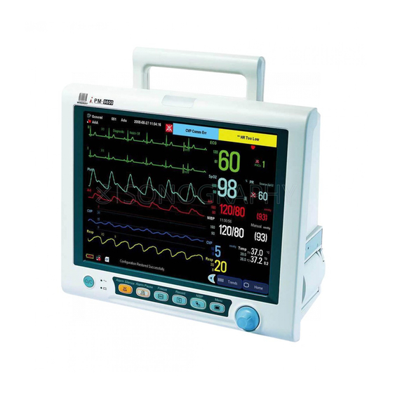
2.3 Display Screen This patient monitor adopts a high-resolution TFT LCD to display patient parameters and waveforms. A typical display screen is shown below. Patient Information Area This area shows the patient information such as department, bed number, patient name, patient category and paced status. - Page 29 Technical Alarm Area This area shows technical alarm messages and prompt messages. When multiple messages come, they will be displayed circularly. Select this area and the technical alarm list will be displayed. Physiological Alarm Area This area shows physiological alarm messages. When multiple alarms occur, they will be displayed circularly.
-
Page 30: Quickkeys
2.4 QuickKeys A QuickKey is a configurable graphical key, located at the bottom of the main screen. They give you fast access to functions. Their availability and the order in which they appear on your screen, depend on how your patient monitor is configured. By default, there are two QuickKeys that remain on the screen all the time to give you fast access to functions. -
Page 31: Basic Operations
WARNING The equipment shall be installed by personnel authorized by Mindray. The software copyright of the equipment is solely owned by Mindray. No organization or individual shall resort to juggling, copying, or exchanging it or to any other infringement on it in any form or by any means without due permission. -
Page 32: Unpacking And Checking
3.1.1 Unpacking and Checking Before unpacking, examine the packing case carefully for signs of damage. If any damage is detected, contact the carrier or us. If the packing case is intact, open the package and remove the equipment and accessories carefully. Check all materials against the packing list and check for any mechanical damage. -
Page 33: Getting Started
3.2 Getting Started 3.2.1 Inspecting the Monitor WARNING Do not use the patient monitor for any monitoring procedure on a patient if you suspect it is not working properly, or if it is mechanically damaged. Contact your service personnel or us. Before you start to make measurements, check the patient monitor for any mechanical damage and make sure that all external cables, plug-ins and accessories are properly connected. -
Page 34: Disconnecting From Power
3.3 Disconnecting from Power To disconnect the patient monitor from the AC power source, follow this procedure: Disconnect the patient cables and sensors from the patient. Press and hold the power on/off switch for above 2 seconds. The patient monitor shuts down and you can unplug the power cable. -
Page 35: Using Keys
3.4.2 Using Keys The monitor has three types of keys: Softkey: A softkey is a graphic key on the screen, giving you fast access to certain menus or functions. The monitor has three types of softkeys: Waveform keys: Each waveform area can be seen as a softkey. You can enter a waveform setup menu by selecting its corresponding waveform area. -
Page 36: Using The Main Menu
3.4.5 Using the Main Menu To enter the main menu, select the hardkey on the monitor’s front. Most of monitor operations and settings can be performed through the main menu. Other menus are similar to the main menu and contain the following parts: Heading: gives a sum-up for the current menu. -
Page 37: Operating Modes
3.5 Operating Modes Your monitor has four operating modes. Some are password protected. Monitoring Mode: This is the normal, every day working mode that you use for monitoring patients. You can change elements such as alarm limits, parameter units and so forth. When you discharge the patient, these elements return to their default values. -
Page 38: Using An External Storage Device
3.6 Using an External Storage Device An external storage device is used to prevent data loss in case of a sudden power failure. The patient data such as trend data, waveform data, etc., will be automatically saved into the external storage device during patient monitoring. In case of a sudden power failure, the patient data can be retrieved from the external storage device after the patient monitor restarts. -
Page 39: Using An External Display
Unload the CF storage card before removing it from the patient monitor. Otherwise it may cause damage to the data in the card. Use only the CF storage card specified by Mindray. 3.7 Using an External Display An external display, showing the same screen as the host display, can be connected to the monitor through the video output connector, for viewing only. -
Page 40: Changing Measurement Settings
3.8.2 Changing Measurement Settings Each measurement has a setup menu in which you can adjust all of its settings. You can enter a setup menu by selecting a certain measurement numeric. For example, by selecting the ECG numeric, you can access the [ECG Setup] menu. This menu displays numeric-related measurement settings, such as alarm limits, alarm switch and so forth. -
Page 41: Setting Up A Monitor
3.9.1 Setting up a Monitor In situations where you install a patient monitor or change the patient monitor’s application site, you need to setup the patient monitor as follows: In the main menu, select [Maintenance >>] → [User Maintenance >>] → enter the required password. -
Page 42: Setting The Date And Time
3.9.5 Setting the Date and Time In the main menu, select [Maintenance >>] → [System Time>>]. Set [Year], [Month], [Day], [Hour], [Minute] and [Second]. To set the date format and the time format, In the main menu, select [Maintenance >>]→[User Maintenance >>]→enter the required password and then select [Device Setup >>]. -
Page 43: Adjusting Volume
3.9.6 Adjusting Volume Alarm Volume In the main menu, select [Alarm Setup >>]. Select the appropriate volume from [Alm Volume]: X-10, in which X is the minimum volume, depending on the set minimum alarm volume (refer to the chapter Alarm), and 10 the maximum volume. - Page 44 FOR YOUR NOTES 3-14...
-
Page 45: Managing Patients
Managing Patients 4.1 Admitting a Patient The patient monitor displays physiological data and stores them in the trends as soon as a patient is connected. This allows you to monitor a patient that is not admitted yet. However, it is recommended that you fully admit a patient so that you can clearly identify your patient, on recordings, reports and networked devices. -
Page 46: Editing Patient Information
WARNING [Patient Cat.] and [Paced] will always contain a value, regardless of whether the patient is fully admitted or not. If you do not specify settings for these fields, the patient monitor uses the default settings from the current configuration, which might not be correct for your patient. -
Page 47: Switching Between Wire And Wireless Networks
NOTE Discharging a patient clears all history data in the monitor. 4.4 Switching between Wire and Wireless Networks To switch between wire and wireless networks: In the main menu, select [Maintenance >>]→ [User Maintenance >>]. Enter the required password. In the user maintenance menu, select [Network Setup] In the network setup menu, select the [Network Type] drop-down box and then toggle between [Wire] and [Wireless]. - Page 48 In the popup [Transfer Patient Data] menu, you can: Select [Transfer Patient] to transfer the patient data to the monitor, or Select [Cancel Transfer] to cancel the operation of transferring patient data. Then the patient data to be transferred becomes archived patient data. The current patient data starts to be saved in the card.
-
Page 49: Connecting To A Central Monitoring System
4.6 Connecting to a Central Monitoring System If your patient monitor is connected to a central monitoring system (CMS): All patient information, measurement data and settings on the patient monitor can be transferred to the CMS. All patient information, measurement data and settings can be displayed simultaneously on the patient monitor and CMS. - Page 50 FOR YOUR NOTES...
-
Page 51: Managing Configurations
Managing Configurations 5.1 Introduction When performing continuous monitoring on a patient, the clinical professional often needs to adjust the monitor’s settings according to the patient’s condition. The collection of all these settings is called a configuration. Allowing you to configure the monitor more efficiently, the monitor offers different sets of configuration to suit different patient categories and departments. -
Page 52: Entering And Exiting The Configuration Mode
Conventional configuration items These items define how the monitor works, e.g., screen layout, record, print and alarm settings. For all the configuration items and their default values, see appendix Configuration Default Information. 5.2 Entering and Exiting the Configuration Mode To enter the configuration mode, Press the hardkey on the monitor’s front to enter the main menu. -
Page 53: Viewing And Changing Configurations
5.3 Viewing and Changing Configurations In configuration mode, you can view, change and save all the configuration items for each department. After entering the configuration mode, verify that the current department is the one you desire. In the system configuration-main screen, select the desired configuration and then select [View Config.]. -
Page 54: Adding A Configuration
NOTE Changing the department will delete all the saved user configurations automatically. Please act with caution. 5.4 Adding a Configuration You can change monitor settings as required and then save the changed settings into a user configuration. You can specify a name for the saved user configuration. The monitor can save up to 3 sets of user configuration for the current department. -
Page 55: Loading A Configuration
In the select user configuration menu, select the user configurations you want to delete and then select [Ok]. Select [Ok] in the popup dialog box. 5.6 Loading a Configuration You may make changes to some settings during operation. However, these changes or the pre-selected configuration may not be appropriate for the newly admitted patient. -
Page 56: Loading The Latest Configuration Automatically
To set the default configuration at startup, Enter the configuration mode. In the load configuration area, select a factory default configuration of the current department or a user configuration as the default configuration. Select [Adu] or [Ped] or [Neo] from the [Patient Cat.] as the default patient category. You can change the department by selecting [Change Department >>]. -
Page 57: Transferring A Configuration
5.9 Transferring a Configuration When installing several monitors with identical user configuration it is not necessary to set each unit separately. An USB storage card may be used to transfer the configuration from monitor to monitor. To export the current monitor’s configuration: Insert the USB storage card into the monitor’s USB port. - Page 58 FOR YOUR NOTES...
-
Page 59: User Screens
User Screens 6.1 Tailoring Your Screens You can tailor your patient monitor’s screens by setting: Waveform sweep mode Wave line size The way to draw waves The color in which each measurement’s numerics and waveform are displayed The waveforms to be displayed and their display order. Some settings can be changed manually in monitoring mode. -
Page 60: Changing Parameter And Waveform Colors
6.1.4 Changing Parameter and Waveform Colors Select [Waveform] in configuration mode. In the [Parameter/Wave Color] area, you can set color for each parameter and waveform. Selecting [Next Page] allows you to set color for more parameters and waveforms. 6.1.5 Selecting Waveforms for Display Your can select waveforms for display on the screen: In the main menu, select [Others >>]→[Wave Setup >>]→[Select Waves >>]. -
Page 61: Viewing Minitrends
6.2 Viewing Minitrends 6.2.1 Having a Split-Screen View of Minitrends You can split the all parameters screen so that one part of the screen, on the left hand side, continuously shows graphic minitrends beside waveforms as shown in the figure below. To have a split-screen view of minitrends, you can: In the main menu, select [Screen Layout >>]→[Minitrends]→[Ok]. -
Page 62: Changing Minitrend Length
6.2.2 Changing Minitrend Length The minitrend length cannot be chosen for individual parameters. You can only change the minitrend length for all parameters: Select either parameter’s minitrend field. In the [Minitrend Setup] menu, select [Minitrend Length] and then select your desired setting. -
Page 63: Viewing Oxycrg
6.3 Viewing oxyCRG To have a split screen view of oxyCRG, you can: In the main menu, select [Screen Layout >>]. Select [oxyCRG]→[Ok]. The split-screen view covers the lower part of the waveform area and shows HR trend, trend and RR trend (or Resp Wave). At the bottom, there are controls: Trend length list box In the trend length list box, you can select [1 min], [2 min], [4 min], or [8 min]. -
Page 64: Viewing Other Patients
6.4 Viewing Other Patients 6.4.1 Care Group If your patient monitor is connected to a central monitoring system, you can select up to 8 bedside monitors into a Care Group. This lets you: View information on the monitor screen from another bed in the same Care Group. Be notified of physiological and technical alarm conditions at the other beds in the same Care Group. -
Page 65: Understanding The View Other Patient Window
6.4.3 Understanding the View Other Patient Window When you first open the [View Other Patient] window, the patient monitor automatically selects a monitor from the network to display in the [View Other Patient] window. The view other patient window covers the lower part of the waveform area and consists of: Information Area: shows the patient information (including department, bed number, patient name, etc.) and network status symbols. -
Page 66: Understanding The Big Numerics Screen
Additionally, you can change a waveform or parameter for viewing. To change a waveform for viewing, select the waveform segment where you want a new waveform to appear and then select the waveform you want from the popup menu. To change a parameter for viewing, select the parameter window where you want a new parameter to appear and then select the parameter you want from the popup menu. -
Page 67: Alarms
Alarms Alarms, triggered by a vital sign that appears abnormal or by technical problems of the patient monitor, are indicated to the user by visual and audible alarm indications. WARNING A potential hazard can exist if different alarm presets are used for the same or similar equipment in any single area, e.g. -
Page 68: Alarm Levels
7.2 Alarm Levels By severity, the patient monitor’s alarms can be classified into three categories: high level, medium level and low level. Physiological alarms Technical alarms Indicate that your patient is in a Indicate a severe device malfunction or an High life threatening situation, such improper operation, which could make it possible... -
Page 69: Alarm Lamp
7.3.1 Alarm Lamp If an alarm occurs, the alarm lamp will flash. The flashing color and frequency match the alarm level as follows: High level alarms: the lamp flashes quickly in red. Medium level alarms: the lamp flashes slowly in yellow. Low level alarms: the lamp turns yellow without flashing. -
Page 70: Reminder Tones
NOTE When multiple alarms of different levels occur simultaneously, the patient monitor will select the alarm of the highest level and give visual and audible alarm indications accordingly. 7.3.5 Reminder Tones When alarms are turned off or alarm tones are paused or turned off, the patient monitor will give a single beep as the reminder tone in case of an active alarm condition. -
Page 71: Switching Off Alarms
When the alarm pause time expires, the alarm paused status is automatically cancelled and the alarm tone will sound. You can also cancel the alarm paused status by pressing the hardkey. You can set the alarm pause time as desired in configuration mode. The default alarm pause time is 2 minutes. -
Page 72: Switching Off Alarm Sound
7.4.4 Switching off Alarm Sound In configuration mode, you can set the minimum alarm volume. The minimum alarm volume refers to the minimum value you can set for the alarm volume. When the alarm volume is set to 0, the monitor stays in alarm sound off status and a symbol appears on the screen. -
Page 73: Displaying Alarm Limits
7.5.2 Displaying Alarm Limits To have a better view of measurement numerics, you can choose not to display alarm limits in the parameter window by switching off displaying alarm limits in configuration mode. For details, refer to the chapter Managing Configuration. 7.5.3 Setting Alarm Delay Time You can set the alarm delay time for over-limit alarms of continuously measured parameters. -
Page 74: Setting The Alarm Level
Setting the Alarm Level Select the parameter window for your desired measurement to enter its setup menu. Select [Alm Lev] and toggle between [High], [Med] and [Low]. You can also set alarm levels for all alarms together: In the main menu, select [Alarm Setup >>]→[Alarm Levels Setup >>]. You can view and set alarm levels for all the ongoing measurements in the popup menu. -
Page 75: Mass Alarm Setup
7.5.5 Mass Alarm Setup In the main menu, select [Alarm Setup >>]→[Mass Alarm Setup>>]. You can review and set alarm limits, alarm switches and alarm recordings for all parameters. 7.5.6 Adjusting Alarm Limits Automatically The monitor can automatically adjust alarm limits according to the measured vital signs, using the auto limits function. -
Page 76: Clearing Technical Alarms
7.7 Clearing Technical Alarms For some technical alarms, their alarm lamp flashing and alarm tones are cleared and the alarm messages change to prompt messages after the hardkey is pressed. After the patient monitor restores the normal alarm status, it can give alarm indications correctly in case these alarms are triggered again. -
Page 77: Using Care Group Alarms
7.10 Using Care Group Alarms 7.10.1 Care Group Auto Alarms When auto alarm is set on for viewing other patient and a Care Group is set up on your monitor, a flashing symbol will appear beside the QuickKeys area if any monitor in your Care Group, which is not currently viewed by your monitor, is alarming. -
Page 78: Silencing Care Group Alarms
7.10.3 Silencing Care Group Alarms You can silence the alarm sound of the currently viewed bed in the view other patient window. This function can be set in configuration mode only. When the alarm silence function for other patients is active and the currently viewed bed is in normal alarm status or alarm sound off status, press the button in the view other patient window. -
Page 79: Monitoring Ecg
Monitoring ECG 8.1 Introduction The electrocardiogram (ECG) measures the electrical activity of the heart and displays it on the patient monitor as a waveform and a numeric. ECG monitoring provides two algorithms: Basic algorithm The Basic algorithm enables 3-, 5- or 12-lead ECG monitoring, ST-segment analysis, arrhythmia analysis and interpretation of resting 12-lead ECG. -
Page 80: Preparing To Monitor Ecg
8.3 Preparing to Monitor ECG 8.3.1 Preparing the Patient and Placing the Electrodes Prepare the patient’s skin. Proper skin preparation is necessary for good signal quality at the electrode, as the skin is a poor conductor of electricity. To properly prepare the skin, choose flat, non-muscular areas and then follow this procedure: Shave hair from skin at chosen sites. -
Page 81: Ecg Lead Placements
8.3.3 ECG Lead Placements The electrode placement illustrations in this chapter adopt the AHA standard. 3-Leadwire Electrode Placement Following is an electrode configuration when using 3 leadwires: RA placement: directly below the clavicle and near the right shoulder. LA placement: directly below the clavicle and near the left shoulder. LL placement: on the left lower abdomen. - Page 82 The chest (V) electrode can be placed on one of the following positions: V1 placement: on the fourth intercostal space at the right sternal border. V2 placement: on the fourth intercostal space at the left sternal border. V3 placement: midway between the V2 and V4 electrode positions. V4 placement: on the fifth intercostal space at the left midclavicular line.
-
Page 83: Switching Ecg Lead Set
WARNING When using electrosurgical units (ESU), patient leads should be placed in a position that is equal distance from the Electrosurgery electrotome and the grounding plate to avoid burns to the patient. Never entangle the ESU cable and the ECG cable together. When using electrosurgical units (ESU), never place ECG electrodes near to the grounding plate of the ESU, as this can cause a lot of interference on the ECG signal. -
Page 84: Checking Paced Status
8.3.5 Checking Paced Status It is important to set the paced status correctly when you start monitoring ECG. The paced symbol is displayed when the [Paced] status is set to [Yes]. The pace pulse markers “ ” are shown on the ECG wave when the patient has a paced signal. To change the paced status, you can select either: the patient information area, or [Patient Setup >>] in the main menu and then [Patient Demographics], or,... -
Page 85: Understanding The Ecg Display
8.4 Understanding the ECG Display In the all parameters screen, the patient monitor allows up to three ECG to be displayed simultaneously. Following is an ECG display screen with 5-lead set and for reference only. Your display may be configured to look slightly different. Lead label of the displayed wave ECG gain ECG filter label... -
Page 86: Changing Ecg Settings
8.5 Changing ECG Settings 8.5.1 Setting Pacemaker Rate (For Mortara Only) Some pacemaker pulses cannot be rejected. When this happens, the pulses are counted as a QRS complex and could result in an incorrect HR and a failure in detecting some arrhythmias. -
Page 87: Changing The Ecg Filter Settings
When [ECG Display] is set to [Normal] and [Sweep Mode] is set to [Refresh], cascaded ECG waveforms can be displayed. To cascade ECG waveforms: Select either ECG wave to enter its lead menu. Select [Cascade] and then select [On]. A cascaded waveform is displayed in two waveform positions. -
Page 88: Switching Defibrillator Synchronization On/Off
Besides, you can set the notch frequency in maintenance mode: In the main menu, Select [Maintenance >>]→[User Maintenance >>]→enter the required password. Select [Device Setup >>] and then set [Notch Filter] to [50Hz] or [60Hz] according to the power line frequency. 8.5.6 Switching Defibrillator Synchronization On/Off As defibrillator synchronization, analog output and nurse call share the same signal output port, you need to set the port for defibrillator synchronization before getting the monitor and... -
Page 89: Selecting Ecg Waves For Display
8.5.7 Selecting ECG Waves for Display In the all parameters screen, the patient monitor allows up to three ECG to be displayed simultaneously. When monitoring with a 5-lead set, you can select [Waveforms] in the ECG setup menu and then select either one, or two, or three waves for display. When you select three ECG waves for display, there must be one ECG wave that will be displayed permanently on the screen. -
Page 90: Adjusting Qrs Volume
For the Mortara algorithm, the system will analyze ECG waveforms from multiple channels simultaneously so as to compute HR and to analyze and detect arrhythmia. 8.5.10 Adjusting QRS Volume When HR is selected as the alarm source, QRS sounds are produced based on the HR. To adjust the QRS volume, select [Beat Vol] in the [ECG Setup] menu and select the appropriate setting. -
Page 91: Changing St Filter Settings
8.6.2 Changing ST Filter Settings When ST-segment analysis is performed, dedicated filters are used to ensure the diagnostic quality. When ST-segment analysis is switched on, [Filter] switches to [Diagnostic] automatically when it is not in the diagnostic mode. 8.6.3 Understanding the ST Display This example shows ST numerics with 5-lead ECG. -
Page 92: Setting St Alarm Delay Time
8.6.6 Setting ST Alarm Delay Time You can set ST alarm delay time by selecting [ST Alarm Delay] from [ST Analysis>>]. The following ST alarm delay time is optional: 30s (default), 45s, 1min, 1.5min, 2 min, and 3min. 8.6.7 Adjusting ST Measurement Points As shown in the figure below, the ST measurement for each beat complex is the vertical difference between two measurement points with the R-wave peak as the baseline for the measurement. -
Page 93: About Arrhythmia Monitoring
Mortara algorithm In the [ST Analysis] menu, select [Adjust ST Points >>]. In the [Adjust ST Points] window, three vertical lines represent the ISO, J and ST points’ positions respectively. Select [View Leads] and use the Knob to select an ECG lead with obvious J point and R wave. -
Page 94: Understanding The Arrhythmia Events
8.7.1 Understanding the Arrhythmia Events Basic algorithm Arrhythmia message Description Asystole No QRS complex for 4 consecutive seconds (in absence of ventricular fibrillation or chaotic signals). Vfib/Vtac A fibrillatory wave for 4 consecutive seconds. A dominant rhythm of adjacent Vs and a HR > the V-Tach Heart Rate Limit. No pace pulse detected for 1.75 x average R-to-R intervals following a QRS complex (for paced patients only). - Page 95 Mortara algorithm Arrhythmia message Description Asystole No QRS complex for 4 consecutive seconds (in absence of ventricular fibrillation or chaotic signals). Vfib Ventricular fibrillation occurs and persists for 6 seconds. Vtac Ventricular HR is greater or equal to the preset threshold and the number of consecutive PVCs is greater than the preset threshold.
-
Page 96: Switching Arrhythmia Analysis On And Off
8.7.2 Switching Arrhythmia Analysis On and Off To switch arrhythmia analysis on or off: Select the ECG parameter window and then select [Arrh. Analysis >>] from the popup menu. Select [Arrh. Analysis] to toggle between [On] and [Off]. PVC numeric When arrhythmia analysis is turned off, the PVC numeric will not be displayed. -
Page 97: Initiating Arrhythmia Relearning Manually
Mortara algorithm Arrh. event Range Default configurations Interval Unit Asys. Delay 2 to 10 NICU: 3 Other departments: 5 Vtac Rate 100 to 200 NICU: 150 Other departments: 130 Vtac PVC 3 to12 beats Multif. PVC’s 3 to 31 beats Window Tachy High Adult: 100 to 300... -
Page 98: Automatic Arrhythmia Relearn
8.7.6 Automatic Arrhythmia Relearn Arrhythmia relearning is initiated automatically whenever: The ECG lead or lead label is changed The HR computing lead is changed. The ECG lead is re-connected A new patient is admitted The paced status setting is changed Arrhythmia analysis is switched on. -
Page 99: 12-Lead Ecg Monitoring
8.8 12-Lead ECG Monitoring 8.8.1 Entering the 12-lead ECG Monitoring Screen Refer to the section 8.3.3 ECG Lead Placement for placing the electrodes. Select the ECG parameter window and then set [Lead Set] to [12-Lead], and [ECG Display] to [12-Lead] in the popup menu. There are totally 12 ECG waves and 1 rhythm wave displayed on the screen. -
Page 100: Interpretation Of Resting 12-Lead Ecg
8.8.2 Interpretation of resting 12-Lead ECG Interpretation of resting 12-lead ECG is intended for adult patients. You can only start an interpretation of resting 12-lead ECG 11 seconds after entering the 12-lead ECG monitoring screen. Otherwise, the prompt message [Not enough data. Cannot analyze.] will be displayed. -
Page 101: Reviewing Interpretation Of Resting 12-Lead Ecg Results
8.8.3 Reviewing Interpretation of resting 12-Lead ECG Results In the 12-lead ECG monitoring screen, you can review previous interpretation of resting 12-lead ECG results by selecting [Review]. In this review window, you can: Switch between [Results] and [Waveforms] for review. Select to view more results. - Page 102 FOR YOUR NOTES 8-24...
-
Page 103: Monitoring Respiration (Resp)
Monitoring Respiration (Resp) 9.1 Introduction Impedance respiration is measured across the thorax. When the patient is breathing, the volume of air changes in the lungs, resulting in impedance changes between the electrodes. Respiration rate (RR) is calculated from these impedance changes, and a respiration waveform appears on the patient monitor screen. -
Page 104: Optimizing Lead Placement For Resp
Lead I Lead II 9.3.1 Optimizing Lead Placement for Resp If you want to measure Resp when you are measuring ECG, you may need to optimize the placement of the two electrodes between which Resp will be measured. Repositioning ECG electrodes from standard positions results in changes in the ECG waveform and may influence ST and arrhythmia interpretation. -
Page 105: Lateral Chest Expansion
9.3.4 Lateral Chest Expansion In clinical applications, some patients (especially neonates) expand their chests laterally, causing a negative intrathoracic pressure. In these cases, it is better to place the two respiration electrodes in the right midaxillary and the left lateral chest areas at the patient’s maximum point of the breathing movement to optimize the respiratory waveform. -
Page 106: Changing Resp Detection Mode
9.5.3 Changing Resp Detection Mode In the [Resp Waveform] menu, select [Detection Mode] and toggle between [Auto] and [Manual]. In auto detection mode, the patient monitor adjusts the detection level automatically, depending on the wave height and the presence of cardiac artifact. Note that in auto detection mode, the detection level (a dotted line) is not displayed on the waveform. -
Page 107: Changing The Size Of The Resp Wave
9.5.4 Changing the Size of the Resp Wave WARNING When monitoring in manual detection mode, make sure to check the respiration detection level after you have increased or decreased the size of the respiration wave. Select the Resp wave to enter the [Resp Waveform] menu. Then, you can select [Gain] and then select an appropriate setting. - Page 108 FOR YOUR NOTES...
-
Page 109: Monitoring Pr
Monitoring PR 10.1 Introduction The pulse numeric counts the arterial pulsations that result from the mechanical activity of the heart. You can display a pulse from any measured SpO or any arterial pressure (see the IBP section). The displayed pulse numeric is color-coded to match its source. Alarm Limits PR Source PR: detected pulsations per minute. -
Page 110: Selecting The Active Alarm Source
10.2.2 Selecting the Active Alarm Source In most cases the HR and PR numerics are identical. In order to avoid simultaneous alarms on HR and Pulse, the monitor uses either HR or Pulse as its active alarm source. To change the alarm source, select [Alm Source] in the [ECG Setup] or [PR Setup] menu and then select either: [HR]: if you want the HR to be the alarm source for HR/PR. -
Page 111: Monitoring Spo
Monitoring SpO 11.1 Introduction monitoring is a non-invasive technique used to measure the amount of oxygenated haemoglobin and pulse rate by measuring the absorption of selected wavelengths of light. The light generated in the probe passes through the tissue and is converted into electrical signals by the photodetector in the probe. -
Page 112: Safety
To identify which SpO module is incorporated into your patient monitor, see the company logo located at the lower left corner on your monitor’s front. If it is: Mindray SpO module, there is no logo. Masimo SpO module, there is the Masimo SET logo. -
Page 113: Changing Spo Settings
When monitoring critically ill patients, selecting shorter averaging time will help understanding the patient’s state. To set the averaging time: For Mindray SpO module, select [Sensitivity] in the [SpO Setup] menu and then toggle between [High], [Med] and [Low], which respectively correspond to 7 s, 9 s and 11 s. -
Page 114: Monitoring Spo 2 And Nibp Simultaneously
11.5.4 Monitoring SpO and NIBP Simultaneously When monitoring SpO and NIBP on the same limb simultaneously, you can switch [NIBP Simul] on in the [SpO Setup] menu to lock the SpO alarm status until the NIBP measurement ends. If you switch [NIBP Simul] off, low perfusion caused by NIBP measurement may lead to inaccurate SpO readings and therefore cause false physiological alarms. -
Page 115: Pitch Tone
After approximately 10.9 seconds, a Sat-Second alarm would sound, because the limit of 50 Sat-Seconds would have been exceeded. Seconds Saturation levels may fluctuate rather than remaining steady for several seconds. Often, the patient % SpO may fluctuate above and below an alarm limit, re-entering the non-alarm range several times. -
Page 116: Measurement Limitations
11.6 Measurement Limitations If you doubt the measured SpO , check patient vital signs first. Then check the patient monitor and SpO sensor. The following factors may influence the accuracy of measurement: Ambient light Physical movement (patient and imposed motion) Diagnostic testing Low perfusion Electromagnetic interference, such as MRI environment... -
Page 117: Nellcor Information
11.8 Nellcor Information Nellcor Patents This device is covered under one or more the following U.S. Patents: 4,802,486; 4,869,254; 4,928,692; 4,934,372; 5,078,136; 5,351,685; 5,485,847; 5,533,507; 5,577,500; 5,803,910; 5,853,364; 5,865,736; 6,083,172; 6,463,310; 6,591,123; 6,708,049; Re.35,122 and international equivalents. U.S.A international patents pending. No Implied License Possession or purchase of this device does not convey any express or implied license to use the device with unauthorized replacement parts which would, alone, or in combination with... - Page 118 FOR YOUR NOTES 11-8...
-
Page 119: Monitoring Nibp
Monitoring NIBP 12.1 Introduction This monitor uses the oscillometric method for measuring the non-invasive blood pressure (NIBP). This measurement can be used for adults, pediatrics and neonates. Automatic non-invasive blood pressure monitoring uses the oscillometric method of measurement. To understand how this method works, we’ll compare it to the auscultative method. -
Page 120: Safety
12.2 Safety WARNING Be sure to select the correct patient category setting for your patient before measurement. Do not apply the higher adult settings for pediatric or neonatal patients. Otherwise it may present a safety hazard. Do not measure NIBP on patients with sickle-cell disease or any condition where skin damage has occurred or is expected. -
Page 121: Setting Up The Nibp Measurement
12.4 Setting Up the NIBP Measurement 12.4.1 Preparing to Measure NIBP Power on the monitor. Verify that the patient category is correct. Change it if necessary. Plug the air tubing into the NIBP connector. Select a correct sized cuff and then apply it as follows: Determine the patient’s limb circumference. -
Page 122: Measurement Methods
12.5 Measurement Methods There are three methods of measuring NIBP: Manual: measurement on demand. Auto: continually repeated measurements at set intervals. STAT: continually rapid series of measurements over a five minute period, then return to the previous mode. 12.5.1 Enabling NIBP Auto Cycling and Setting the Interval Select the NIBP parameter window to enter the [NIBP Setup] menu. -
Page 123: Understanding The Nibp Numerics
12.6 Understanding the NIBP Numerics The NIBP display shows numerics only as below. Your display may be configured to look slightly different. Time of last measurement Time remaining to next measurement Measurement mode Unit of pressure: mmHg or kPa Alarm Limits Prompt message area: shows NIBP-related prompt messages Systolic pressure Mean pressure... -
Page 124: Changing Nibp Settings
12.7 Changing NIBP Settings 12.7.1 Choosing NIBP Alarm Source You can monitor for alarm conditions in systolic, diastolic and mean pressure, either singly or in parallel. In the [NIBP Setup], select [Alm Source] and choose from: [Sys]: alarms are given only when the systolic pressure violates the alarm limits. [Dia]: alarms are given only when the diastolic pressure violates the alarm limits. -
Page 125: Setting The Cuff Inflation Pressure
12.7.4 Setting the cuff inflation pressure You can set the initialcuff inflation pressure . In the [NIBP Setup] menu, select [Inflation Pressure] and then select the appropriate setting. 12.8 Assisting Venous Puncture You can use the NIBP cuff to cause sub-diastolic pressure to block the venous blood vessel and therefore help venous puncture. -
Page 126: Nibp Accuracy Test
Follow this procedure to perform the leakage test: Set the patient category to [Adu]. Connect the cuff to the NIBP connector on the monitor. Wrap the cuff around the cylinder as shown below. Cylinder Monitor Connector for Air tubing Cuff NIBP cuff In the main menu, select [Maintenance >>]→[User Maintenance >>]→enter the required password and then select [Maintain NIBP]→[NIBP Leakage Test]. -
Page 127: Calibrating Nibp
Follow this procedure to perform the accuracy test: Connect the equipment as shown. Monitor Manometer Tubing Connector for NIBP cuff Balloon pump Metal vessel Before inflation, the reading of the manometer should be 0.If not, disconnect the airway and reconnect it until the readings is 0. In the main menu, select [Maintenance >>]→[User Maintenance >>]→enter the required password and then select [Maintain NIBP]→[NIBP Accuracy Test]. - Page 128 FOR YOUR NOTES 12-10...
-
Page 129: Monitoring Temp
Monitoring Temp 13.1 Introduction This patient monitor allows you to monitor two temperature sites simultaneously and calculate the temperature difference between them. 13.2 Safety WARNING Verify that the probe detection program works correctly before monitoring. Besides, make sure that if you plug out the temperature probe cable from the T1 or T2 connector, the monitor will trigger a technical alarm and give the alarm message [T1 Sensor Off] or [T2 Sensor Off] correctly. -
Page 130: Understanding The Temp Display
13.4 Understanding the Temp Display The temperature monitoring is displayed on the monitor as three numerics: T1, T2 and TD. By selecting this area, you can enter the [Temp Setup] menu. 13.5 Setting the Temperature Unit In the [Temp Setup] menu, select [Unit] and toggle between [ºC] and [ºF]. 13-2... -
Page 131: Monitoring Ibp
Monitoring IBP 14.1 Introduction The monitor can monitor up to 4 invasive blood pressures and displays the systolic, diastolic and mean pressures and a waveform for each pressure. 14.2 Safety WARNING Use only pressure transducers specified in this manual. Never reuse disposable pressure transducers. - Page 132 Connect the pressure line to the patient catheter. Position the transducer so that it is level with the heart, approximately at the level of the midaxillary line. Select the appropriate label. Zero the transducer. After a successful zeroing, turn off the stopcock to the atmosphere and turn on the stopcock to the patient.
-
Page 133: Understanding The Ibp Display
14.4 Understanding the IBP Display The IBP measurement is displayed on the monitor as a waveform and numeric pressures. The figure below shows the waveform and numerics for the Art pressure. For different pressures, this display may be slightly different. Waveform Systolic pressure Diastolic pressure... -
Page 134: Changing Ibp Settings
14.5 Changing IBP Settings 14.5.1 Changing a Pressure for Monitoring Select the pressure you want to change to enter its setup menu. Select [Label] and then select your desired label from the list. The already displayed labels cannot be selected. Label Description Label... -
Page 135: Changing Averaging Time
14.5.3 Changing Averaging Time The IBP value displayed on the monitor screen is the average of data collected within a specific time. The shorter the averaging time is, the quicker the patient monitor responds to changes in the patient’s blood pressure. Contrarily, the longer the averaging time is, the slower the patient monitor responds to changes in the patient’s blood pressure, but the measurement accuracy will be improved. -
Page 136: Measuring Pawp
14.6 Measuring PAWP Pulmonary Artery Wedge Pressure (PAWP) values, used to assess cardiac function, are affected by fluid status, myocardial contractility, and valve and pulmonary circulation integrity. Obtain the measurement by introducing a balloon-tipped pulmonary artery flotation catheter into the pulmonary artery. When the catheter is in one of the smaller pulmonary arteries, the inflated balloon occludes the artery allowing the monitor to record changes in the intrathoracic pressures that occur throughout the respiration cycle. - Page 137 NOTE After entering the PAWP measurement window, the monitor will turn off the PA alarm automatically. 14-7...
-
Page 138: Setting Up The Pawp Measurement
14.6.2 Setting Up the PAWP Measurement Wedge the flotation catheter into the pulmonary artery. Then inflate the balloon and pay attention to PA waveform changes on the screen. After obtaining a stable PAWP waveform, press the [Freeze] key to freeze the waveform and deflate the balloon. -
Page 139: Zeroing The Transducer
14.7 Zeroing the Transducer To avoid inaccurate pressure readings, the monitor requires a valid zero. Zero the transducer in accordance with your hospital policy (at least once per day).Zero whenever: A new transducer or adapter cable is used. You reconnect the transducer cable to the monitor. The monitor restarts. - Page 140 FOR YOUR NOTES 14-10...
-
Page 141: Monitoring Cardiac Output
Monitoring Cardiac Output 15.1 Introduction The cardiac output (C.O.) measurement invasively measures cardiac output and other hemodynamic parameters using the right heart (atria) thermodilution method. A cold solution of known volume and temperature is injected into the right atrium through the proximal port of a pulmonary artery (PA) catheter. -
Page 142: Influencing Factors
15.3 Influencing Factors The factors that affect cardiac output are: temperature of injectate solution, volume of injectate solution, patient’s baseline blood temperature, patient’s inspiratory/expiratory cycle, The degree of the catheter’s distal end in close proximity to the lung, the catheter itself, the patient rhythm and hemodynamic status, and any other rapid IV solutions which are infused while the C.O. - Page 143 Injectate C.O. cable Patient monitor PA catheter Syringe Stopcock Hole In-line probe Balloon inflation port Ice water Check if the height and weight are appropriate for your patient in the [Patient Demographics] menu. Change if necessary. 4. Select the C.O. parameter window to enter the [C.O. Setup] menu. In the [C.O. Setup] menu: Check that the correct computation constant is entered.
- Page 144 A. Currently measured numeric Currently measured C.O. curve Prompt message area D. Buttons Averaged values Measurement windows When you see the message [Ready for New Measurement], select the [Start] button and then inject the solution within 4 seconds. As shown in the figure above, during the measurement, the currently measured thermodilution curve is displayed.
-
Page 145: Measuring The Blood Temperature
In the buttons area, you can: Select [Start] to start a C.O. measurement. Select [Stop] to stop the current measurement. Select [Cancel] during a measurement to cancel the measurement. Selecting it after a measurement deletes the measured results. Select [X-Scale] to adjust the scale of the X-axis. Options for scale range are 30 s and 60 s. -
Page 146: Changing C.o. Settings
15.6 Changing C.O. Settings 15.6.1 Setting the Temperature Unit In the [C.O. Setup] menu, select [Temp Unit] to toggle between [ºC] and [ºF]. 15.6.2 Setting the Interval between Measurements To avoid inaccurate measurements, a certain period of time should be allowed for the blood temperature to become stable before starting a new measurement. -
Page 147: Monitoring Carbon Dioxide
Monitoring Carbon Dioxide 16.1 Introduction monitoring is a continuous, non-invasive technique for determining the concentration of in the patient’ airway by measuring the absorption of infrared (IR) light of specific wavelengths. The CO has its own absorption characteristic and the amount of light passing the gas probe depends on the concentration of the measured CO When a specific band of IR light is passed through respiratory gas samples, some of IR light will be absorbed by the CO... -
Page 148: Preparing To Measure Co
16.2 Preparing to Measure CO 16.2.1 Using a Sidestream CO Module Attach the watertrap to the module and then connect the CO components as shown below. Watertrap fixer Sampling line Watertrap By default, the sidestream CO module is in standby mode. Access the [CO Setup] menu to set [Operating Mode] to [Measure]. - Page 149 CAUTION The watertrap collects water drops condensed in the sampling line and therefore prevents them from entering the module. If the collected water reaches a certain amount, you should drain it to avoid blocking the airway. The watertrap has a filter preventing bacterium, water and secretions from entering the module.
- Page 150 16.2.3 Using a Mainstream CO module Connect the sensor to the module. By default, the microstream CO module is in measure mode. The message [CO Sensor Warmup] appears on the screen when the CO module is plugged. After warm-up is finished, connect the transducer to the airway adapter. Perform a zero calibration per the Zeroing the Sensor section.
-
Page 151: Entering The Standby Mode
16.3 Changing CO Settings 16.3.1 Entering the Standby Mode The standby mode of the CO module relates to the standby mode of the monitor as follows: If the monitor enters the standby mode, the CO module will also enter the standby mode. -
Page 152: Setting Up Humidity Compensation
For the microstream CO module, gas compensations are not required. For the maintream CO module, in the [CO Setup] menu, respectively select: [Balance Gas] and toggle between [Room Air], [N O] and [He]. Select [Room Air] when air predominates in the ventilation gas mixture, [N O] when N O predominates in the ventilation gas mixture, and [He] when He predominates in the ventilation gas... -
Page 153: Setting The Apnea Alarm Delay
16.3.5 Setting the Apnea Alarm Delay In the [CO Setup] menu, select [Adjust CO Limits >>] → [Apnea Time] and then select the appropriate setting. The monitor will alarm if the patient has stopped breathing for longer than the preset apnea time. 16.3.6 Choosing a Time Interval for Peak-Picking For microstream and mainstream CO modules, you can select a time interval for picking the... -
Page 154: Setting Barometric Pressure Compensation
16.4 Setting Barometric Pressure Compensation The mainstream CO module does not have the function of automatic barometric pressure compensation. For the mainstream CO module, the default barometric pressure is 760 mmHg. You must modify the barometric pressure based on the actual situation as follows: In the main menu, select [Maintenance >>] →... -
Page 155: Removing Exhaust Gases From The System
16.7 Removing Exhaust Gases from the System WARNING Anesthetics: When using the Sidestream or Microstream CO measurement on patients who are receiving or have recently received anesthetics, connect the outlet to a scavenging system, or to the anesthesia machine/ventilator, to avoid exposing medical staff to anesthetics. -
Page 156: Calibrating The Sensor
After warm-up is finished, connect the sensor to a clean, dry airway adapter. The adapter should be vented to the air and isolated from CO sources, such as ventilator, the patient’s breathing, your own breathing, etc. Select [Start Zero Cal.] in the [CO Setup] menu. -
Page 157: Monitoring Ag
Monitoring AG 17.1 Introduction The anesthetic gas (AG) module measures the patient’s anesthetic and respiratory gases, and can be integrated into the optional O module. The AG module determines the concentration of certain gases using the infrared (IR) light absorption measurement. The gases that can be measured by the AG module absorb IR light. Each gas has its own absorption characteristic. -
Page 158: Preparing To Measure Ag
WARNING To avoid explosion hazard, do not use flammable anesthetic agent such as ether and cyclopropane for this equipment. NOTE The AG module is configured with automatic barometric pressure compensation function. 17.2 Preparing to Measure AG Select an appropriate watertrap according to patient category and attach it to the module. Connect the gas sample line to the connector of the watertrap. - Page 159 CAUTION Position the airway adapter so that the part connecting to the gas sample line is pointing upwards. This prevents condensed water from passing into the gas sample line and causing an occlusion. The watertrap collects water drops condensed in the sampling line and therefore prevents them from entering the module.
-
Page 160: Understanding The Ag Display
17.3 Understanding the AG Display The AG module can send waves and numerics for all measured anesthetic gases for display on the monitor, including: O and AA waves awRR: airway respiratory rate MAC: minimal alveolar concentration End tidal (Et) and fraction of inspired (Fi) numerics for CO O and AA The patient monitor can display up to four anesthetic gas waveforms simultaneously, which are CO... -
Page 161: Mac Values
17.4 MAC Values Minimum alveolar concentration (MAC) is the minimum concentration of the agent in the alveoli. It is a basic index to indicate the depth of anesthesia. The standard ISO 21647 defines MAC as this: alveolar concentration of an inhaled anesthetic agent that, in the absence of other anesthetic agents and at equilibrium, prevents 50% of patients from moving in response to a standard surgical stimulus. -
Page 162: Changing Ag Settings
17.5 Changing AG Settings 17.5.1 Selecting an Anesthetic Gas for Monitoring As the AION 02-type module cannot automatically identify 5 anesthetic gases, you need to select an anesthetic gas before use. To select the anesthetic gas, in the [Gas Setup] menu, select [Agent] and then select the appropriate setting. -
Page 163: Entering The Standby Mode
17.5.6 Entering the Standby Mode For the AG module, the default operating mode is measure. When you set the AG module to the standby mode, the AG gas sample intake pump automatically sets the sample flow rate to zero. When exiting the standby mode, the AG module continues to work at preset sample flow rate with no need to warm up again. -
Page 164: Measurement Limitations
The AION 03-type AG module can identify two anesthetic agents automatically. When the proportion of the primary and secondary anesthetic agents in the mixture changes and the secondary anesthetic agent becomes dominant, the primary and seconday anesthetic agents will be exchanged for display and a physiological alarm may be triggered. 17.7 Measurement Limitations The following factors may influence the accuracy of measurement: Leaks or internal venting of sampled gas... -
Page 165: Removing Exhaust Gases From The System
17.9 Removing Exhaust Gases from the System WARNING Anesthetics: When using the AG measurement on patients who are receiving or have recently received anesthetics, connect the outlet to a scavenging system, or to the anesthesia machine/ventilator, to avoid exposing medical staff to anesthetics. To remove the sample gas to a scavenging system, connect an exhaust tube to the gas outlet connector of the module. - Page 166 FOR YOUR NOTES 17-10...
-
Page 167: Freezing Waveforms
Freezing Waveforms During patient monitoring, the freeze feature allows you to freeze the currently displayed waveforms on the screen so that you can have a close examination of the patient’s status. Besides, you can select any frozen waveform for recording. 18.1 Freezing Waveforms To freeze waveforms, select the hardkey on the monitor’s front,... -
Page 168: Unfreezing Waveforms
18.3 Unfreezing Waveforms To unfreeze the frozen waveforms, you can either: Select [Exit] in the [Freeze] menu, Select the hardkey on the monitor’s front, or Perform any operation through which the screen will re-arrange or a menu will pop up, such as plugging in or out a module, pressing the hardkey, etc. -
Page 169: Review
Review 19.1 Accessing Respective Review Windows In the main menu, select [Review >>]. Select [Graphic Trends], [Tabular Trends], [NIBP], [Arrh. Events], [Alarms], [Full Disclosure] or [12-Lead Analysis] to access their respective review windows. For details about reviewing arrhythmia events and interpretation of resting 12-lead ECG results, refer to the chapter Monitoring ECG. -
Page 170: Reviewing Graphic Trends
19.2 Reviewing Graphic Trends In the [Review] menu, select [Graphic Trends] to access the following window. A. Parameter combo box Slider Parameter value D. Parameter scale Graphic trends Time axis In this review window: To select parameters for viewing, you can either: Select respective parameter combo boxes and then select your desired parameters, Select [Trend Group] and then select a group from the popup list, or Select [Select Parameter>>] and then select your desired parameters from the... - Page 171 You can change the resolution of the trend data by selecting [Interval] and then selecting the appropriate setting: [1 s]: select to view up to 1 hours of graphic trends at 1-second resolution. [5 s]: select to view up to 8 hours of graphic trends at 5-second resolution. [1 min], [5 min] or [10 min]: select to view up to 96 hours of graphic trends at 1-, 5-, or 10-minute resolution.
-
Page 172: Reviewing Tabular Trends
19.3 Reviewing Tabular Trends In the [Review] menu, select [Tabular Trends] to access the following window. In this review window: To select parameters for viewing, you can either: Select [Trend Group] and then select a group from the popup list, or Select the [Define Group>>] button and then select the parameters you want to view from the popup menu. - Page 173 You can change the resolution of the trend data by selecting [Interval] and then selecting the appropriate setting: [5 s] or [30 s]: select to view up to 8 hours of tabular trends at 5- or 30-second resolution. [1 min], [5 min], [10 min], [30 min] or [1 h]: select to view up to 96 hours of tabular trends at your selected resolution.
-
Page 174: Reviewing Nibp Measurements
19.4 Reviewing NIBP Measurements In the [Review] menu, select [NIBP] to access the following window. This window displays systolic pressure, diastolic pressure, mean pressure, pulse rate and time for each measurement. Among them, pulse rate comes from NIBP measurements. Besides, the total number of measurements is displayed at the lower left corner. -
Page 175: Reviewing Alarms
19.5 Reviewing Alarms When a measurement alarm occurs, all relative measurement numerics at the alarm time and related waveforms 2, 4 or 8 seconds respectively before and after the alarm trigger time are stored. To review the stored alarms, select [Review]→[Alarms] to access the following window. -
Page 176: Reviewing Waveforms
You can select beside [Scroll] to navigate through the waveforms. By selecting the [Record] button, you can print out the currently displayed alarm events by the recorder. By selecting the [Print] button, you can print out the currently displayed alarm events by the printer. - Page 177 In this review window: Before reviewing full-disclosure waveforms, your monitor must be equipped with an external storage card (only the CF storage card is supported so far) and you need to save waveforms first. To review full-disclosure waveforms, select [Save Waves >>] and then select the parameters whose waveforms you want to view.
- Page 178 FOR YOUR NOTES 19-10...
-
Page 179: Calculations
Calculations 20.1 Introduction The calculation feature is available with your patient monitor. The calculated values, which are not directly measured, are computed based on the values you provide. Your can perform the following calculations: Dose Calculations Oxygenation Calculations Ventilation Calculations Hemodynamic Calculations Renal Calculations To perform a calculation, select [Calc >>] in the main menu or the [Calc] QuickKey and... -
Page 180: Dose Calculations
20.2 Dose Calculations 20.2.1 Performing Calculations In the main menu, select [Calc >>]→[Dose >>]. Select, in turn, [Patient Cat.] and [Drug Name] and then select the appropriate settings. The dose calculation program has a library of commonly used drugs, of which Drug A through Drug E are for those not specified in this library. -
Page 181: Titration Table
20.2.3 Titration Table To open the titration table, select [Titration Table >>] in the dose calculation window after the dose calculation is finished. In the titration table, when you change: [Reference] [Interval] [Dose Type] The titrated values change accordingly. You can also: Select , or beside the vertical scrollbar to view more values. -
Page 182: Entered Parameters
20.3.2 Entered Parameters Abbreviation Unit Full spelling C.O. L/min cardiac output percentage fraction of inspired oxygen mmHg partial pressure of oxygen in the arteries PaCO mmHg partial pressure of carbon dioxide in the arteries arterial oxygen saturation mmHg partial pressure of oxygen in venous blood venous oxygen saturation hemoglobin ml/L... -
Page 183: Ventilation Calculations
20.4 Ventilation Calculations 20.4.1 Performing Calculations In the main menu, select [Calc >>]→[Ventilation >>]. Enter values for calculation. Select the [Calculate] button. The system performs a calculation per the current settings and displays the calculated values. The values that exceed the range are displayed in yellow background. You can select [Range] to view its normal range in the unit field. -
Page 184: Calculated Parameters
20.4.3 Calculated Parameters Abbreviation Unit Full spelling mmHg partial pressure of oxygen in the alveoli AaDO mmHg alveolar-arterial oxygen difference Pa/FiO mmHg oxygenation ratio a/AO arterial to alveolar oxygen ratio L/min minute volume volume of physiological dead space Vd/Vt physiologic dead space in percent of tidal volume L/min alveolar volume 20.5 Hemodynamic Calculations... -
Page 185: Entered Parameters
20.5.2 Entered Parameters Abbreviation Unit Full spelling C.O. L/min cardiac output heart rate PAWP mmHg pulmonary artery wedge pressure Art Mean mmHg artery mean pressure PA Mean mmHg pulmonary artery mean pressure mmHg central venous pressure end-diastolic volume Height height Weight weight 20.5.3 Calculated Parameters... -
Page 186: Renal Calculations
20.6 Renal Calculations 20.6.1 Performing Calculations In the main menu, select [Calc >>]→[Renal >>]. Enter values for calculation. Select the [Calculate] button. The system performs a calculation per the current settings and displays the calculated values. The values that exceed the range are displayed in yellow background. You can select [Range] to view its normal range in the unit field. -
Page 187: Calculated Parameters
20.6.3 Calculated Parameters Abbreviation Unit Full spelling URNaEx mmol/24h urine sodium excretion URKEx mmol/24h urine potassium excretion Na/K sodium potassium ratio ml/24h clearance of sodium Clcr ml/min creatinine clearance rate FENa fractional excretion of sodium Cosm ml/min osmolar clearance ml/h free water clearance U/P osm None... - Page 188 FOR YOUR NOTES 20-10...
-
Page 189: Recording
Recording 21.1 Using a Recorder The thermal recorder records patient information, measurement numerics, up to three waveforms, etc. Start/Stop key: press to start a recording or stop the current recording. Indicator On: when the recorder works correctly. Off: when the monitor is switched off. Flashes: if an error occurred to the recorder, e.g., the recorder runs out of paper. -
Page 190: Overview Of Recording Types
21.2 Overview of Recording Types By the way recordings are triggered, the recordings can be classified into the following categories: Manually-triggered realtime recordings. Timed recordings. Alarm recordings triggered by an alarm limit violation or an arrhythmia event. Manually-triggered, task-related recordings. The task-related recordings include: Frozen wave recording Graphic trends recording... -
Page 191: Starting And Stopping Recordings
21.3 Starting and Stopping Recordings To manually start a recording, you can either: Select the hardkey on the front of either the patient monitor or the recorder module, Select the [Record] button from the current menu or window. Automatic recordings will be triggered in the following conditions: Timed recordings will start automatically at preset intervals. -
Page 192: Setting The Realtime Recording Length
21.4.3 Setting the Realtime Recording Length The recording time depends on your monitor’s settings when a realtime recording is started. In the [Record Setup] menu, select [Realtime Rec. Length] and toggle between [8 s] and [Continuous]. [8 s]: record 8-second waveforms from the current moment. [Continuous]: record the waveforms from the current moment until stopped manually. -
Page 193: Removing Paper Jam
Paper roll CAUTION Use only specified thermal paper. Otherwise, it may cause damage to the recorder’s printhead, the recorder may be unable to print, or poor print quality may result. Never pull the recorder paper with force when a recording is in process. Otherwise, it may cause damage to the recorder. -
Page 194: Cleaning The Recorder Printhead
21.7 Cleaning the Recorder Printhead If the recorder has been used for a long time, deposits of paper debris may collect on the printhead compromising the print quality and shortening the lifetime of the roller. Follow this procedure to clean the printhead: Take measures against the static electricity such as Disposable Wrist Strap for the work. -
Page 195: Printing
Printing 22.1 Printer The monitor can output patient reports via a connected printer. So far, the monitor supports the following printer: HP LaserJet 1606N HP LaserJet 2035N HP LaserJet 4015N HP LaserJet 1505N The specifications of the reports the monitor prints are: Paper: A4, Letter Resolution: 300 dpi Print on One/Both Sides: printing on one and both sides are supported if the printer... -
Page 196: Starting Report Printouts
Select a connected printer Select [Printer] and then select a connected printer as the monitor’s printer. Search for a printer If your selected printer is not in the list or a new printer is added into the network, you can select the [Search Printer] to re-search for all printers in the network. Set up the paper Select [Paper Size] and toggle between [A4] and [Letter]. -
Page 197: Stopping Report Printouts
22.5 Stopping Report Printouts To stop report printouts, select[Print Setup >>]→[Stop All Reports] in the main menu. 22.6 Setting Up Reports 22.6.1 Setting Up ECG Reports You can print out ECG reports only under full-screen, half-screen or 12-lead monitoring screen. To set up ECG reports, select [Print Setup >>]→[ECG Reports >>] in the main menu. -
Page 198: Setting Up Graphic Trends Reports
[Select Parameter >>]: from the popup menu, you can: [Currently Displayed Trended Parameters]: print the parameter trend data selected from the [Tabular Trends]. [Standard Parameter Group]: select the standard parameter group for printing. [Custom]: You can define a parameter group for printing from the parameters displayed in the low part of the menu. -
Page 199: Printer Statuses
22.8 Printer Statuses 22.8.1 Printer Out of Paper When the printer runs out of paper, the print request will not be responded. If there are too many print jobs that are not responded, a printer error may occur. In these cases, you need to install paper and then re-send the print request. - Page 200 FOR YOUR NOTES 22-6...
-
Page 201: Other Functions
Other Functions 23.1 Marking Events During patient monitoring, some events may exert effects on the patient and as a result change the waveforms or numerics displayed on the monitor. To help analyzing the waveforms or numerics at that time, you can mark these events. To mark an event, Select [Mark Event] QuickKey, or select [Mark Event >>] in the main menu. -
Page 202: Transferring Data
23.3 Transferring Data You can transfer the patient data saved in the monitor to a PC via a crossover network cable or an external storage card, or within a LAN for data management, review or print. 23.3.1 Data Export System You must install the data export system on the intended PC before performing the data transfer operation. -
Page 203: Nurse Call
Transfer data within a LAN Before transferring data within a LAN, do as follows: Connect the patient monitor and the intended PC into the same LAN and acquire the PC’s IP address. Make sure that the data export system is active on the PC. Follow the same procedure as via a crossover network cable to transfer data. - Page 204 Select [Contact Type] and toggle between [Normally Open] and [Normally Closed]. [Normally Open]: select if your hospital’s nurse call relay contact is normally open. [Normally Closed]: select if your hospital’s nurse call relay contact is normally closed. Another two options, [Alm Lev] and [Alarm Cat.], must be set in configuration mode. [Alm Lev] defines the alarm level for nurse call-triggering alarms, and [Alarm Cat.] defines the category within which the nurse call-triggering alarms fall.
-
Page 205: Wireless Network
23.5 Wireless Network The patient monitors, each equipped with a wireless network card, constitute a wireless network via AP (access point).The designated service engineer or personnel shall be responsible for installing and configuring the wireless network for you and perform relative performance tests as well. - Page 206 FOR YOUR NOTES 23-6...
-
Page 207: Batteries
Batteries 24.1 Overview This monitor is designed to operate on battery (sealed lead-acid or Lithium Ion battery) power during intra-hospital patient transfer or whenever the power supply is interrupted. The battery is charged automatically when the monitor is connected to AC power, no matter the monitor is powered on or not. -
Page 208: Installing Or Replacing A Battery
24.2 Installing or Replacing a Battery The patient monitor uses two battery packs. One battery pack can be easily exchanged while the monitor operates from the other. If the patient monitor uses one battery pack, you should insert a new battery pack before the old one depletes. To install or replace a battery, follow this procedure: Open the battery door. -
Page 209: Battery Guidelines
24.3 Battery Guidelines Life expectancy of a battery depends on how frequent and how long it is used. For a properly maintained and stored lead-acid or lithium ion battery, its life expectancy is about 2 or 3 years respectively. For more aggressive use models, life expectancy can be less. We recommend replacing lead acid batteries every 2 years and lithium ion batteries every 3 years. -
Page 210: Battery Maintenance
24.4 Battery Maintenance Conditioning a Battery A battery should be conditioned before it is used for the first time. A battery conditioning cycle is one uninterrupted charge of the battery, followed by an uninterrupted battery discharge and charge. . Batteries should be conditioned regularly to maintain their useful life. -
Page 211: Battery Recycling
Checking a Battery The battery performance test must be performed every two years, before monitor repairs, or whenever the battery is suspected as being the source of the problems.The performance of a rechargeable battery may deteriorate over time. To check the performance of a battery, follow this procedure: Disconnect the monitor from the patient and stop all monitoring or measuring. - Page 212 FOR YOUR NOTES 24-6...
-
Page 213: Care And Cleaning
Care and Cleaning Use only the substances approved by us and methods listed in this chapter to clean or disinfect your equipment. Warranty does not cover damage caused by unapproved substances or methods. We make no claims regarding the efficacy of the listed chemicals or methods as a means for controlling infection. -
Page 214: Cleaning
25.2 Cleaning Your equipment should be cleaned on a regular basis. If there is heavy pollution or lots of dust and sand in your place, the equipment should be cleaned more frequently. Before cleaning the equipment, consult your hospital’s regulations for cleaning the equipment. Recommended cleaning agents are: mild soap (diluted) ammonia (diluted) -
Page 215: Maintenance
Maintenance WARNING Failure on the part of the responsible individual hospital or institution employing the use of this equipment to implement a satisfactory maintenance schedule may cause undue equipment failure and possible health hazards. The safety checks or maintenance involving any disassembly of the equipment should be performed by professional servicing personnel. -
Page 216: Service Tasks
26.2 Service Tasks The following tasks are for our qualified service professionals only. Contact a qualified service provider if your patient monitor needs the following services. Clean and disinfect equipment to decontaminate it before testing or maintaining it. Maintenance schedule Frequency At least once every two years, after any repairs where the Safety checks according to... -
Page 217: Calibrating Ecg
26.4 Calibrating ECG The ECG signal may be inaccurate due to hardware or software problems. As a result, the ECG wave amplitude becomes greater or smaller. In that case, you need to calibrate the ECG module. In the main menu, select [Maintenance >>]→[User Maintenance >>]→enter the required password and then select [Calibrate ECG] from the popup menu. -
Page 218: Calibrating Co2
26.6 Calibrating CO2 For sidestream and microstream CO2 modules, a calibration is needed every year or when the measured values have a great deviation. For maintream CO2 module, no calibration is needed. Tools required: Gas bottle, with 4%, 5% or 6% of CO2. T-shape connector Tubing Follow this procedure to perform a calibration:... -
Page 219: Calibrating Ag
26.7 Calibrating AG Calibrate the AG module every year or when the measured value has a great deviation. Tools required: Gas bottle, with a certain standard gas or mixture gas. Gas concentration should meet the following requirements: AA>1.5%, CO2>1.5%, N2O>40%, O2>40%, of which AA represents an anesthetic agent. -
Page 220: Setting Up Ip Address
Enter the vented gas concentration. If you use only one gas for calibration, set other gases’ concentration to 0. Select [Start] to start calibration. If the calibration is finished successfully, the message [Calibration Completed!] is displayed. If the calibration failed, the message [AG Cal. Failed] is displayed. Perform another calibration. -
Page 221: Accessories
Accessories WARNING Use accessories specified in this chapter. Using other accessories may cause damage to the patient monitor or not meet the claimed specifications. Single-use accessories are not designed to be reused. Reuse may cause a risk of contamination and affect the measurement accuracy. Check the accessories and their packages for any sign of damage. - Page 222 Cable Sets 3-Electrode Cable Sets Compatible Type Model Patient Category Length Part No. Remark with EL6302A 0.6 m 0010-30-42725 / Adult, pediatric EL6304A 0010-30-42732 Long EL6306A Neonate 0.6 m 0010-30-42897 / EL6308A Pediatric 0.6 m 0010-30-42899 / Clip EL6301A 0.6 m 0010-30-42726 / Adult, pediatric EL6303A...
-
Page 223: Spo2 Accessories
0010-20-42712 Sensors The SpO sensor materials that patients or other staff will come into contact with have undertaken the bio-compatibility test and are verified to be in compliance with ISO 10993-1. Mindray SpO Module Type Model Patient Category Length Part No. - Page 224 Pediatric, infant (finger type) 9000-10-07308 OXI-A/N Adult, neonate (finger type) 9000-10-07336 Wavelength emiited by the sensors intended for Mindray SpO module: red right: 660 nm, infrared light: 905 nm. Wavelength emitted by the sensors intended for Masimo SpO module: red light: 660 nm, infrared light: 940 nm.
-
Page 225: Nibp Accessories
27.3 NIBP Accessories Tubing Type Patient Category Length Part No. Adult, pediatric 6200-30-09688 Reusable Neonate 1.5 m 6200-30-11560 Reusable Cuff Patient Measurement Limb Circumference Bladder Model Part No. Category Site (cm) Width (cm) CM1201 Infant 10 to 19 0010-30-12157 CM1202 Pediatric 18 to 26 12.2 0010-30-12158... -
Page 226: Temp Accessories
27.4 Temp Accessories Extension Cable Type Model Temp probe Length Part No. Reusable MR420B MR411, MR412 0011-30-37391 Temp Probes Measurement Type Model Patient Category Length Part No. Site MR401B Esophageal/Rectal 0011-30-37392 Adult MR403B Skin 3.8 m 0011-30-37393 Reusable MR402B Esophageal/Rectal 0011-30-37394 Pediatric, neonate MR404B... - Page 227 ICP Model Material Part No. Gaeltec TYPE.S13 12Pin ICP cable 0010-30-42742 Gaeltec ICT/B Intracranial Pressure Transducer 6800-30-50759 Intracranial Pressure Transducer (global) 0010-10-12151 It is proved through tests that the following accessories are compatible with the patient monitor. Only the accessories proceeded by “*” are available from our company. If you want to purchase other accessories, contact respective manufacturers and make sure if these accessories are approved for sale in local.
-
Page 228: C.o. Accessories
M02A-10-25938 Cannula DRYLINE Airway Adult, pediatric, Straight 9000-10-07486 Adapter neonate Airway adapter: 2 Adult/pediatric watertrap: 2 Mindray CO2 Adult/pediatric Sampling line: 2 accessories package Adult, pediatric Adult Nasal CO2 Sample 6800-30-50618 (M02A) Cannula: 2 Pediatric Nasal CO2 Sample Cannula: 2... - Page 229 Microstream CO Module Disposable Airway Sampling Line Model Patient Category Remark Part No. XS04620 0010-10-42560 XS04624 Humidified 0010-10-42561 Adult, pediatric 7768 Long 0010-10-42563 7737 Long, humidified 0010-10-42564 6324 Humidified 0010-10-42562 Infant, Neonate 7738 Long, humidified 0010-10-42565 Disposable Nasal Sampling Line Model Patient Category Remark...
- Page 230 Mainstream CO Module Material Model Patient Category Remark Part No. 6421 Airway Adult Disposable, with mouthpiece 0010-10-42663 adapter 9960STD 0010-10-42670 Adult Mask 9960LGE Large 0010-10-42671 9960PED Pediatric 0010-10-42669 Cable management 0010-10-42667 straps Sensor 0010-10-42668 holding clips Adult, pediatric, Sensor Capnostat5 Reusable 6800-30-50760 neonate...
-
Page 231: Ag Accessories
27.8 AG Accessories Material Patient Category Remark Part No. Adult, pediatric 9200-10-10530 Watertrap Reusable Neonate 9200-10-10574 Adult, pediatric 9200-10-10533 Sampling line Disposable Neonate 9200-10-10555 Airway Adult, pediatric, neonate Disposable, straight 9000-10-07486 adapter Adult, pediatric, neonate Disposable, elbow 9000-10-07487 Straight airway adapter:2 AG module Elbow airway adapter:2 accessories... -
Page 232: Others
27.9 Others Material Part No. Lead-acid battery M05-302R3R--- M05-010001-06 (black) Lithium battery 0146-00-0099 (gray) 3-core power cord 509B-10-05996 3-core power cord (V3203C-V1625) 0000-10-10903 Grounding cable 1000-21-00122 Defibrillator synchronization cable 6800-20-50781 Nurse call cable 8000-21-10361 CF storage card 023-000067-00 Recorder TR6F-30-67306 Recorder paper (48 mm) A30-000001--- Recorder for update (installed on the device) -
Page 233: Product Specifications
Product Specifications A.1 Monitor Safety Specifications A.1.1 Classifications The patient monitor is classified, according to IEC60601-1: Classification Name Specifications Class I equipment, with both internal and external power supply Type of protection When you doubt the integrity of the external protective earth (ground) against electric shock or the protective earth conductor, switch the equipment to the internal power supply (batteries). - Page 234 A.1.2 Environmental Specifications Operating conditions 0 to 40 5 to 40 (with Sidestream CO module) Temperature (ºC) 0 to 40 (with Microstream CO module) 10 to 40 (with Mainstream CO module or AG module) Relative humidity 15% to 95% (noncondensing) 428.6-808.3 mmHg(57.0-107.5 kPa)...
- Page 235 A.1.3 Power requirements Parameter Specifications AC Power Supply Specifications Line Voltage 100 to 240 ~ Current 1.3 to 0.5 A Frequency 50/60 Hz Fuse T3.15 A H, 250 V Internal battery Number of batteries Type Sealed lead-acid battery or lithium-ion battery Time to shutdown >5 min (after the first low-power alarm) Sealed lead-acid battery...
- Page 236 A.2 Hardware specifications A.2.1 Physical Specifications Size 320×270×180mm (width×height×depth) Weight Standard configuration: <5.0kg (without batteries and recorder) A.2.2 Display Host display Screen type Colour TFT LCD Screen Size (diagonal) 12.1" Resolution 800×600 pixels A.2.3 Recorder Method Thermal dot array Horizontal resolution 16 dots/mm (25 mm/s paper speed) Vertical resolution 8 dots/mm...
- Page 237 A.2.5 Audio Indicator Give alarm tones (45 to 85 dB), key tones, QRS tones; support Speaker PITCH TONE and multi-level tone modulation; alarm tones comply with IEC60601-1-8. A.2.6 Alarm Signal Alarm output delay Less than 1 second Pause duration 1, 2, 3, 5, 10, 15 minutes or infinite, depending on configuration A.2.7 Monitor Interface Specifications Power 1 AC power input connector...
- Page 238 A.2.8 Signal Output Auxiliary Output Meets the requirements of EC60601-1 for short-circuit Standard protection and leakage current ECG Analog Output Diagnostic mode: 0.05 to 150 Hz Bandwidth (-3dB; reference Monitor mode: 0.5 to 40 Hz frequency:10Hz) Surgical mode: 1 to 20 Hz Max transmission delay 25 ms (in diagnostic mode, and with Notch off) Sensitivity...
- Page 239 A.3 Data Storage Trends: 96 hours, at 1 min resolution Trends Minitrends: 1 hour, at 1 s resolution 100 alarms and related parameter waveforms. The waveform Parameter alarms recording length can be 8s, 16s or 32s. 80 arrhythmia events and relate waveforms. The waveform Arrh.
- Page 240 A.5.1 ECG Meet standards of EC11, EC13, EN60601-2-27/IEC60601-2-27, Standards IEC60601-2-25, YY91079, GB9706.25 3-lead: I, II, III 5-lead: I, II, III, aVR, aVL, aVF, V Lead set 12-lead: I, II, III, aVR, aVL, aVF, V1 to V6 ECG standard AHA, IEC 1.25 mm/mV (X0.125), 2.5 mm/mV (X0.25), 5 mm/mV (X0.5), Display sensitivity 10 mm/mV (X1), 20 mm/mV (X2), 40mm/mV (X4), Auto...
- Page 241 Pace Pulse Pace pulses meeting the following conditions are labelled with a PACE marker: Pace pulse markers Amplitude: ±2 to ±700 mV Width: 0.1 to 2 ms Rise time: 10 to 100 µs When tested in accordance with the ANSI/AAMI EC13: Sections 4.1.4.1 and 4.1.4.3, the heart rate meter rejects all pulses meeting the following conditions.
- Page 242 In compliance with the requirements in Clause 4.1.2.1 d) of ANSI/AAMI EC13, the following method is used: If the last 3 consecutive RR intervals are greater than 1200 ms, the 4 HR averaging method most recent RR intervals are averaged to compute the HR. Otherwise, heart rate is computed by subtracting the maximum and minimum ones from the most recent 12 RR intervals and then averaging them.
- Page 243 Mortara algorithm Only the differences from the Basic algorithm are listed. In compliance with the requirements in Clause 4.1.2.1 d) of ANSI/AAMI EC13, the following method is used: Heart rate is computed by averaging the most recent 16 RR HR averaging method intervals, unless the HR by averaging the most recent 4 heart beats is less than or equals to 48 bpm.
- Page 244 (low limit + 1) to 100 SpO2 Low Desat to (high limit – 1) Desat 50 to (high limit – 1) Mindray SpO Module Standards Meet standards of ISO9919 *Measurement accuracy verification: The SpO2 accuracy has been verified in human experiments by comparing with arterial blood sample reference measured with a CO-oximeter.
- Page 245 Sensor type Totally neonates Data Arms 518B 97 (51 male & 46 female) 200 pairs 2.38% 520N 122 (65 male & 57 female) 200 pairs 2.88% The Pulse Oximeter with neonatal SpO2 sensors was also validated on adult subjects. Refreshing rate 7 s (When the sensitivity is set to High) averaging time 9 s (When the sensitivity is set to Medium)
- Page 246 A.5.4 PR Alarm limit Range (bpm) Step (bpm) PR High (low limit +2) to 240 PR Low 25 to (high limit-2) PR from Mindray SpO Module Measurement range 20 to 254 bpm Resolution 1 bpm Accuracy ±3 bpm Refreshing rate...
- Page 247 PR from Nellcor SpO Module Measurement range 20 to 300 bpm Resolution 1 bpm 20 to 250 bpm: ±3 bpm Accuracy 251 to 300 bpm, not specified Refreshing rate SPO2 averaging time 8 s, 16 s PR from IBP Module Measurement range 25 to 350 bpm Resolution...
- Page 248 A.5.5 NIBP Meet standards of EN60601-2-30/IEC60601-2-30, EN1060-1, Standards EN1060-3, EN1060-4 and SP10 Technique Oscillometry Mode of operation Manual, Auto and STAT Auto mode repetition intervals 1, 2, 3, 4, 5, 10, 15, 30, 60, 90, 120, 180, 240 or 480 min Typical measurement time 20 to 40 s (depending on the heart rate and motion artifact) STAT mode cycle time...
- Page 249 Alarm limit Range (mmHg) Step (mmHg) Adult: (low limit+5) to 270 Sys High Pediatric: (low limit+5) to 200 Neonate: (low limit+5) to 135 Sys Low 40 to (high limit-5) Adult: (low limit+5) to 230 Mean High Pediatric: (low limit+5) to 165 Neonate: (low limit+5) to 110 Mean Low 20 to (high limit-5)
- Page 250 A.5.6 Temp Standards Meet standard of EN12470-4 Technique Thermal resistance 0 to 50 °C (32 to 122 °F) Measurement range 0.1 °C Resolution ±0.1 °C or ±0.2 °F (without probe) Accuracy Refreshing rate Body surface: <100 s Time for accurate measurement Body cavity: <80 s Alarm limit...
- Page 251 Alarm limit Range (mmHg) Step (mmHg) Sys High Mean High (low limit + 2) to 300 Dia High Sys Low 0 to (high limit – 2) Mean Low Dia Low Sys High Mean High (low limit + 2) to 120 Dia High Sys Low Mean Low...
- Page 252 Alarm limit Range Step (low limit + 1) to 43 °C TB High (low limit + 1.8) to 109.4 °F 0.1 °C 0.1 °F 23 to (high limit - 1) °C TB Low 73.4 to (high limit - 1.8) °F A.5.9 CO Standard Meet standard of ISO 21647...
- Page 253 Measured with a neonatal watertrap and a 2.5-meter adult sampling line: <4 s @ 150 ml/min <5 s @ 120 ml/min <5 s @ 100 ml/min <6.5 s @ 70 ml/min awRR measurement range 0 to 120 rpm awRR measurement precision ±2 rpm Apnea time 10 s, 15 s, 20 s, 25 s, 30 s, 35 s, 40 s...
- Page 254 Microstream CO Module Measurement range 0 to 99 mmHg 0 to 38 mmHg: ±2 mmHg Accuracy* 39 to 99 mmHg: ±5% of the reading+0.08% of (the reading-38) Accuracy drift Meet the requirement for measurement accuracy within 6 hours * Accuracy applies for respiration rate up to 80 rpm. For respiration rate above 80 rpm, the accuracy is 4 mmHg or ±12% of the reading, whichever is greater.
- Page 255 Mainstream CO Module Measurement range 0 to 150 mmHg 0 to 40 mmHg: ±2 mmHg 41 to 70 mmHg: ±5% of the reading Accuracy 71 to 100 mmHg: ±8% of the reading 101 to 150 mmHg: ±10% of the reading Accuracy drift Meet the requirement for measurement accuracy within 6 hours 0 to 69 mmHg:...
- Page 256 ±0.3% Iso accuracy ±(8% Other anesthetic gases: Gases Range (% Accuracy (% 0 to 1 ±0.1 1 to 5 ±0.2 5 to 7 ±0.3 7 to 10 ±0.5 >10 Not specified 0 to 20 ±2 20 to 100 ±3 0 to 25 ±1 25 to 80 ±2...
- Page 257 gas sample flow rate 120ml/min, using the DRYLINE™ watertrap and neonatal DRYLINE™ sampling line (2.5m): ≤250 ms (fall time: 200ms) ≤250 ms ≤600 ms Hal, Iso, Sev, Des ≤300 ms ≤350 ms Rise time (10 % to 90%) gas sample flow rate 200ml/min, using the DRYLINE™ water trap and adult DRYLINE™...
- Page 258 Effect of interference gases on AG measurements Quantitive effect (% Concentration (% ) Agent 1) 2) Agent Nitrogen ≤78% Xenon <100% Helium <50% Ethanol <0.1% Acetone <1% Methane <1% Saturated Isopropanol vapour Metered dose inhaler Unspecified Unspecified Unspecified 0.5 propellants, Methoxyflurane Unspecified Unspecified Unspecified Unspecified 1) Agent represents one of Des, Iso, Enf, Sev, and Hal.
- Page 259 Alarm limit Range Step EtCO2 High (low limit + 2) to 76 mmHg EtCO2 Low 0 to (high limit - 2)mmHg 1 mmHg FiCO2 High (low limit + 2) to 76 mmHg FiCO2 Low 0 to (high limit - 2)mmHg awRR High (low limit + 2) to 100 rpm 1 rpm...
- Page 260 FOR YOUR NOTES A-28...
- Page 261 The equipment meets the requirements of EN 60601-1-2. NOTE Use of accessories, transducers, and cables other than those specified may result in increased emission and/or decreased immunity of the equipment. The equipment should not be used adjacent to or stacked with other equipment, and if adjacent or stacked use is necessary, the equipment should be observed to verify normal operation in the configuration in which it will be used.
- Page 262 TABLE 1 Guidance and declaration — electromagnetic emissions The equipment is intended for use in the electromagnetic environment specified below. The customer or the user of the equipment should assure that it is used in such an environment. Electromagnetic environment — guidance Emissions test Compliance RF emissions...
- Page 263 TABLE 2 Guidance and declaration — electromagnetic immunity The equipment is intended for use in the electromagnetic environment specified below. The customer or the user of the equipment should assure that it is used in such an environment. Immunity test IEC 60601 Test Compliance Electromagnetic environment...
- Page 264 TABLE 3 Guidance and declaration — electromagnetic immunity The equipment is intended for use in the electromagnetic environment specified below. The customer or the user of the equipment should assure that it is used in such an environment Immunity IEC 60601 Compliance Electromagnetic environment —...
- Page 265 TABLE 4 Recommended separation distances between portable and mobile RF communication and the equipment The equipment is intended for use in an electromagnetic environment in which radiated RF disturbance are controlled. The customer or the user of the equipment can help prevent electromagnetic interference by maintaining a minimum distance between portable and mobile RF communication equipment (transmitters) and the equipment as recommended below, according to the maximum output power of the communication equipment.
- Page 266 FOR YOUR NOTES...
- Page 267 Default Configurations This chapter lists some of the most important factory default settings for each department in configuration mode. You cannot change the factory default configuration itself. However, you can make changes to the settings from the factory default configuration and then save the changed configuration as a user configuration.
- Page 268 C.2 Parameters Configuration C.2.1 ECG ECG Setup Item Name General NICU User Defaults Lead Set * * 5-Lead 3-Lead 5-Lead ECG Waveforms * * 2 Alm Source * * HR Alarm * * On Alm Lev * * Med HR High HR Low Sweep * * 25 mm/s...
- Page 269 ST Analysis Item Name Algorithm General NICU User Defaults ST Unit ST Analysis / Alarm Alm Lev ST-X High / when ST Unit is mV: 0.20 when ST Unit is mm: ST-X Low when ST Unit is mV: -0.20 when ST Unit is mm: -2.0 Basic 78 ms...
- Page 270 Arrh. Analysis Item Name Algorithm Genral OR ICU NICU CCU User Defaults Arrh. Analysis PVCs Alarm Alm Lev PVCs High Arrhythmia Threshold Settings Asystole Mortara Vtac Rate Vtac PVCs Multif. PVC’s Window Tachy Adu: Ped: Neo: Brady Adu: Ped: Neo: Arrhythmia Alarm Settings X Alarm Basic...
- Page 271 C.2.2 RESP Item Name General NICU User Defaults Alarm Alm Lev Sweep 6.25 mm/s Lead Gain Resp High Adu, Ped: Neo: Resp Low Adu, Ped: Neo: Apnea Time 20 s 15 s 20 s Detection Mode Auto C.2.3 PR Item Name General OR NICU User Defaults...
- Page 272 C.2.4 SpO2 Item Name General ICU NICU CCU User Defaults Alarm Alm Lev SpO2 High Adu, Ped: Neo: SpO2 Low Adu, Ped, Neo: Desat Limit Adu, Ped, Neo: Sweep 25 mm/s NIBP Simul Pitch Tone Mode 1 Sensivity Sensivity (Masimo) Normal Averaging (Masimo, Nellcor) Sat-Seconds (Nellcor)
- Page 273 C.2.6 NIBP Item Name General NICU User Defaults Alarm * * On Alm Lev * * Med Alm Source * * All Measure Mode * * Manual Auto Manual Auto Interval * * 15 min 5 min 15 min 30 min 15 min Press.
- Page 274 C.2.7 IBP Item Name General OR NICU User Defaults Alarm Alm Lev P1 Measure P2 Measure Mean P3 Measure P4 Measure Mean Alarm Source Press. Unit mmHg Response Sweep 25 mm/s Auto Scaling Filter 12.5 Hz Wave Overlapping Art, Ao, UAP, BAP, FAP Setup Upper Scale Middle Scale Lower Scale...
- Page 275 PA Setup Upper Scale Middle Scale Lower Scale Sys High (mmHg) Sys Low (mmHg) Mean High (mmHg) Mean Low (mmHg) Dia High (mmHg) Dia Low (mmHg) CVP, LAP, RAP, ICP, UVP Setup Upper Scale Middle Scale Lower Scale Mean High (mmHg) Mean Low (mmHg)
- Page 276 C.2.8 C.O. Item Name General OR NICU User Defaults Alarm Alm Lev TB High (ºC) 39.0 TB Low (ºC) 36.0 Comp. Const 0.542 Auto Ti (ºC) Manual Ti (ºC) Temp Unit ºC Interval (s) C.2.9 CO2 Item Name General NICU User Defaults Alarm * * On...
- Page 277 Item Name General OR ICU NICU User Defaults Microstream CO2 Setup Humidity Compen Max Hold 20 s Auto Standby (min) Mainstream CO2 Setup Max Hold 10 s O2 Compen Balance Gas Room Air AG Compen Alarm Limits Setup EtCO2 High (mmHg) * * Adu, Ped: Neo: EtCO2 Low (mmHg)
- Page 278 C.2.10 AG Item Name General NICU CCU User Defaults Alarm Alm Lev Sweep 6.25 mm/s O2 Compen Operating Mode Measure Flow Rate Adu, Ped: 120 ml/min Neo: 70 ml/min Auto Standby Apnea Time 20 s CO2 Setup CO2 Unit mmHg Wave Type Draw Scale...
- Page 279 Gas Setup Agent O2 Unit N2O Scale O2 Scale when unit is mmHg: when unit is % or KPa: AA Scale Hal/Enf/Iso Scale Des Scale Sev Scale EtO2 High EtO2 Low FiO2 High FiO2 Low EtN2O High EtN2O Low FiN2O High FiN2O Low EtHal/Enf/Iso High EtHal/Enf/Iso Low...
- Page 280 C.3 Routine Configuration C.3.1 Alarm Item Name General OR ICU NICU CCU User Defaults Alarm Pause Time 2 min Minimum Alarm Volume Alm Volume Reminder Tone Reminder Vol Reminder Interval 1 min Latching Alarms Display Alarm Limits Alm Lev of ECG Lead Off Alm Lev of SpO2 Sensor Off * Alarm Tone Interval High Level Alarm:...
- Page 281 C.3.2 Screens Item Name General OR ICU NICU CCU User Defaults Select Screen All parameters Select Wave Sequence for All Pleth Parameters IBP1 Screen IBP2 IBP3 IBP4 Select Parameter 1 Parameters for Parameter 2 SpO2 Big Numerics Parameter 3 Resp Screen Parameter 4 NIBP...
- Page 282 Purple Orange Cyan Yellow RESP Yellow Yellow Blue Green Orange Purple Cyan Yellow C.O. White * X represents a waveform label, such as ECG, RESP, CO2 and so forth. The ECG waveform cannot be set off. C.3.4 Archives Item Name General NICU User Defaults...
- Page 283 C.3.5 Record Item Name General OR NICU User Defaults Waveform 1 ECG1 Waveform 2 ECG2 Waveform 3 Length Interval Paper Speed 25 mm/s Gridlines Alm Rec * X represents a parameter label. C.3.6 Print Item Name General NICU CCU User Defaults Printer * None...
- Page 284 Reports Interval * * Auto Select Parameters * * Currently Displayed Trended Parameters Realtime Set as End Case Report * * Unselected Report Sweep * * Auto Select Wave * * Current C.3.7 Others Item Name General ICU NICU CCU User Defaults Brightness * * 5...
-
Page 285: Alarm Messages
Alarm Messages This chapter lists only the most important physiological and technical alarm messages. Some messages appearing on your monitor may not be included. In this chapter: The “I” field indicates how alarm indications are cleared: “A” means all alarm indications can be cleared, “B”... - Page 286 D.1 Physiological Alarm Messages Measurement Alarm messages Cause and solution XX Too High XX value has risen above the high alarm limit or fallen below the low alarm limit. Check the patient’s XX Too Low condition and check if the patient category and alarm limit settings are correct.
- Page 287 Measurement Alarm messages Cause and solution CO2 Apnea The patient stops breathing, or the respiration signal was so weak that the monitor cannot perform AG Apnea respiration analysis. Check the patient’s condition and the RM connections. FiO2 Too Low Check the patient’s condition, the ventilated O content and the AG connections.
- Page 288 Measurement Alarm message Cause and solution ECG Artifact Artifacts are detected on the ECG analysis lead and as a result heart rate cannot be calculated and Asystole, Vfib and Vtac cannot be analyzed. Check the connections of the electrodes and leadwires and check for any possible source of interference around the cable and electrode.
- Page 289 Measurement Alarm message Cause and solution the patient’s condition and change the sensor SpO2 Weak Signal application site. If the error persists, replace the SpO2 Weak Pulse sensor. SpO2 Low Perf SpO2 Interference The SpO2 signal has been interfered. Check for any possible sources of signal noise around the sensor and check the patient for great motion.
- Page 290 Measurement Alarm message Cause and solution NIBP Illegally Reset An illegal reset occurred during NIBP measurement. Check if the airway is occluded. YYSensor Off Check the sensor connection and reconnect the sensor. (YY represents an IBP label.) C.O. TB Sensor Off CO2 Sensor High Check, stop using or replace the sensor.
- Page 291 Measurement Alarm message Cause and solution CO2 Check Sensor The CO2 sensor appears abnormal. Check or replace the sensor. CO2 No Sensor Make sure that the CO2 sensor is connected. CO2 Temp The module detects that the temperature has Overrange exceeded the specified range.
- Page 292 Measurement Alarm message Cause and solution allow the batteries to charge. Battery Depleted Battery Mismatched The two batteries have different charge capacity, or the batteries unspecified have been used, or there is a problem with the batteries. Make sure that correct batteries are used and the batteries are not damaged, or replace the batteries.
- Page 293 Symbols and Abbreviations E.1 Symbols µA microampere µV microvolt ampere ampere hour beat per minute bit per second ºC centigrade cubic centimeter centimeter decibel dyne second ºF fahrenheit gram gigahertz gutta hour hertz inch kilogram kilopascal litre pound meter milliampere hour mega byte microgram milli-equivalents...
- Page 294 millimeter mmHg millimeters of mercury centimeters of water millisecond millivolt milliwatt MΩ megaohm nanometer breaths per minute second volt volt ampere Ω watt – minus, negative percent per; divide; or plus equal to < less than > greater than ≤ less than or equal to ≥...
- Page 295 E.2 Abbreviations AaDO alveolar-arterial oxygen gradient AAMI Association for Advancement of Medical Instrumentation alternating current adult anaesthesia gas American Heart Association ANSI American National Standard Institute aortic pressure arterial left foot augmented lead left arm augmented lead right arm augmented lead awRR airway respiratory rate brachial aterial pressure...
- Page 296 direct current desflurane diastolic dot per inch electrocardiograph end-diastolic volume European Economic Community electromagnetic compatibility electromagnetic interference enflurane electrosurgical unit end-tidal EtCO end-tidal carbon dioxide end-tidal nitrous oxide ethylene oxide end-tidal oxygen femoral arterial pressure Federal Communication Commission Food and Drug Administration fraction of inspired FiCO fraction of inspired carbon oxygen...
- Page 297 internet protocol isoflurane left arm left atrial pressure lateral liquid crystal display left cardiac work LCWI left cardiac work index light emitting diode left leg LVDS low voltage differential signal LVSW left ventricular stroke work LVSWI left ventricular stroke work index minimum alveolar concentration mean arterial pressure Medical Device Directive...
- Page 298 right right arm random access memory right atrial pressure record, recording Resp respiration reduced hemoglobin right leg respiration rate arterial oxygen saturation sevoflurane self-maintenance stroke index arterial oxygen saturation from pulse oximetry systolic time ratio stroke volume systemic vascular resistance SVRI systemic vascular resistance index Sync...
- Page 300 PN: 9211-20-87439(7.0)...
Need help?
Do you have a question about the iPM-9800 and is the answer not in the manual?
Questions and answers
Срок службы монитора ipm-9800
The document does not specify the overall lifespan of the Mindray iPM-9800 monitor. However, it mentions that lead acid batteries should be replaced every 2 years and lithium-ion batteries every 3 years, with battery performance tests recommended every two years.
This answer is automatically generated