Table of Contents
Advertisement
Advertisement
Chapters
Table of Contents

Related Manuals for Vatech PaX-i3D


Summary of Contents for Vatech PaX-i3D
- Page 1 (PHT-60CFO) User Manual Version 1.5.0 E n g l i s h...
-
Page 5: Notice
PANO, CEPH, and 3D CT imaging capabilities into a single system. This manual describes how to operate the PaX-i3D Green system. It is recommended that you thoroughly familiarize yourself with this manual in order to make the most effective use of this equipment. -
Page 6: Table Of Contents
3.1.2 Features ................26 3.1.3 The PaX-i3D Green Model Series (Optional)......28 3.1.4 Standards and Regulations...........29 Imaging System Structure ........... 30 General View of the PaX-i3D Green ........31 3.3.1 Control Panel ................33 3.3.2 Touch Screen ................34 3.3.3 Emergency Stop Switch ............39 3.3.4... - Page 7 PANO ..................50 4.3.2 CEPH ...................55 4.3.3 CBCT ...................57 Getting Started ..........64 Turning on the PaX-i3D Green ..........64 Running the Image Viewer ..........65 5.2.1 Creating a New Patient Record ..........65 5.2.2 Retrieving Patient Records ...........67 Initiating the Imaging Program ..........69 Acquiring PANO Images ........
- Page 8 10.2.2 Maintenance Task Checklis ..........132 Disposing of the Unit ........134 Technical Specifications ......136 12.1 Mechanical Specifications ..........136 12.2 Technical Specifications ............ 140 12.3 Electrical Specifications ............. 145 12.4 Environmental Specifications ..........145 PaX-i3D Green (PHT-60CFO) User Manual...
- Page 9 Age group: classification table .......... 161 Positioning the pediatric dental patient ......161 Setting exposure values to the age group ......162 The references pertinent to the potential risks for the pediatric patients ................162 Abbreviations ..........164 PaX-i3D Green (PHT-60CFO) User Manual...
-
Page 11: General Information
General Information Manufacturer's Liability ............12 Owner and Operator's Obligations ........12 Conventions in this Manual ..........13... -
Page 12: Manufacturer's Liability
■ Genuine VATECH approved equipment and components have been used at all times. ■ All maintenance and repairs have been performed by a VATECH authorized agent. ■ The equipment has been used normally in accordance with the user's manual. -
Page 13: Conventions In This Manual
IMPORTANT potentially cause problems to the equipment and/or its operation. IMPORTANT Emphasizes important information or provide NOTE useful tips and hints. NOTE Indicates a component which must be replaced SINGLE USE for each new patient. PaX-i3D Green (PHT-60CFO) User Manual... -
Page 15: Warnings And Precautions
Warnings and Precautions General Safety Precautions ..........16 Electricity-related Safety Precautions ........20 Radiation Safety ..............21 Warnings ................22... -
Page 16: General Safety Precautions
■ Do not push or pull the equipment. If equipment overbalances, resulting in the risk of physical injuries or property damage. ■ Operators must ask the patient to remain still until the equipment arm has stopped moving and the reset motion is complete. PaX-i3D Green (PHT-60CFO) User Manual... - Page 17 ■ If this product is exposed to water, moisture, or a foreign substance, immediately turn off all power to the equipment and contact your VATECH technical support representative. ■ Immediately cease all operation of this equipment if there are signs of oil leakage and contact you VATECH technical support representative.
- Page 18 ■ Do not position the patient in the equipment while it is initiating as the patient could be injured if the equipment malfunctions. ■ Ensure that the patient is kept clear of the mechanism while adjusting the height of the equipment. PaX-i3D Green (PHT-60CFO) User Manual...
- Page 19 ■ No part of this equipment is serviceable by the user. All maintenance and repair of this equipment must be performed by a qualified service technician. ■ This product may only be operated with original VATECH accessories or third-party accessories expressly approved by VATECH. PaX-i3D Green (PHT-60CFO) User Manual...
-
Page 20: Electricity-Related Safety Precautions
5 m of the unit, please observe the specifications stated in the installation requirements. Portable and mobile RF communications equipment may interfere with ■ medical electrical equipment. Therefore, the use of mobile wireless phones in medical offices or hospital environments must be prohibited. PaX-i3D Green (PHT-60CFO) User Manual... -
Page 21: Radiation Safety
■ The operator must immediately stop imaging if the equipment malfunctions. As a manufacturer of radiology equipment that conforms to stringent protection standards around the world, we guarantee the maximum degree of protection against WARNING radiation hazards for our equipment. PaX-i3D Green (PHT-60CFO) User Manual... -
Page 22: Warnings
Dental X-ray system. WARNING ● PaX-i3D Green system is designed to withstand the effects of defibrillation. However, when possible, disconnect the Dental X-ray systems during defibrillation since a malfunction of the safety controls could otherwise result in electrical burns for the patient. - Page 23 ■ If a problem occurs during imaging, press the red emergency stop button to immediately stop all moving parts and cut off all power to the equipment’s electrical components. ■ Never touch the patient while also touching the SIP/SOP connectors. PaX-i3D Green (PHT-60CFO) User Manual...
- Page 24 ■ If the equipment is not stable, property damage and/or personal injury may occur. ■ Do not push or pull the equipment. ■ Equipment should only be installed by an authorized technician, complying with proper installation procedures. PaX-i3D Green (PHT-60CFO) User Manual...
-
Page 25: Pax-I3D Green Imaging System Overview
3.1.2 Features ................26 3.1.3 The PaX-i3D Green Model Series (Optional)......28 3.1.4 Standards and Regulations...........29 Imaging System Structure ........... 30 General View of the PaX-i3D Green ........31 3.3.1 Control Panel ................33 3.3.2 Touch Screen ................34 3.3.3 Emergency Stop Switch ............39 3.3.4... -
Page 26: Introduction
2D and 3D of the head region, including the dental/maxillofacial regions. The PaX-i3D Green may only be used by dentists, X-ray technicians and other professionals who are licensed to perform X-rays by the law of the region in which it is being used. - Page 27 3 PaX-i3D Green Imaging System Overview ■ Fast CEPH: CEPH imaging with low X-ray dose and high speed ■ Control Panel: to allow an easy handling of the equipment ■ Multi-FOV (Field Of View): FOV is selected, based on the most suitable ROI.
-
Page 28: The Pax-I3D Green Model Series (Optional)
3 PaX-i3D Green Imaging System Overview 3.1.3 The PaX-i3D Green Model Series (Optional) Model Item Sensor FOV (mm) PANO Xmaru1501CF Xmaru1215CF MP 100 X 80, 80 X 80, 80 X 50, 50 X 50 PANO+CBCT 160 X 100, 120 X 90, 80 X 80, 80 X 50, 50 X 50... -
Page 29: Standards And Regulations
3 PaX-i3D Green Imaging System Overview 3.1.4 Standards and Regulations Standards: The PaX-i3D Green is designed and manufactured to meet the following standards: IEC/EN/UL 60601-1, IEC/EN 60601-1-1, IEC/EN 60601-1-2, IEC/EN 60601-1- 3, IEC/EN 60601-2-7, IEC/EN 60601-2-28, IEC/EN 60601-2-32, IEC/EN 60601- 2-44, ISO 9001, ISO 13485 NEMA standard publication PS 3.1-3.18, 2008... -
Page 30: Imaging System Structure
3 PaX-i3D Green Imaging System Overview Imaging System Structure RS232 Interface Cable 10 m (32.8’) 1. Signal input/output at the column up/down switch (optional) External equipment intended for connection to signal input, signal output or other connectors, shall comply with relevant IEC Standard (e.g., IEC60950 for IT equipment... -
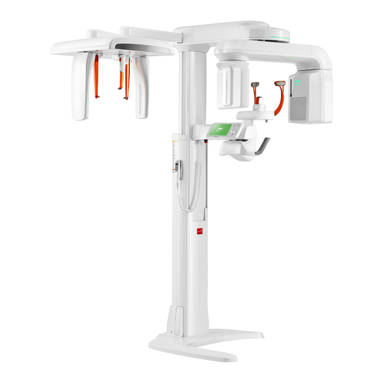
Page 31: General View Of The Pax-I3D Green
3 PaX-i3D Green Imaging System Overview General View of the PaX-i3D Green PaX-i3D Green (PHT-60CFO) User Manual... - Page 32 The portion of the column fixed to the base. BASE Used to balance and stabilize the equipment. The structural overview of the PaX-i3D Green may differ depending on the model. For more information, refer to NOTE section “3.1.3 The PaX-i3D Green Model Series”.
-
Page 33: Control Panel
3 PaX-i3D Green Imaging System Overview 3.3.1 Control Panel Buttons Functions Configures the parameter settings in each Touch Screen imaging mode. For more information on this, refer to 3.3.2 Touch Screen. Laser Beam ON / OFF Turns ON or OFF the laser beams. -
Page 34: Touch Screen
3 PaX-i3D Green Imaging System Overview 3.3.2 Touch Screen You can configure the parameter settings in each mode using the touch screen. The touch screen on the equipment and the imaging program (See 4.3 Imaging Software Overview) on the PC are synchronized in real time, and display the same parameter settings. - Page 35 3 PaX-i3D Green Imaging System Overview Function Description Examination area selection Selects the tooth position Guidance Image INFO Function Tube voltage UP / DOWN Tube current UP / DOWN Patient’s gender selection Patient’s X-ray intensity selection Displays the parameter settings...
- Page 36 3 PaX-i3D Green Imaging System Overview Temple Support Adjustment Button Touch Screen LOCK Touch Screen is automatically locked while exposure. RETURN Returns the Rotating Unit back to its initial position. PaX-i3D Green (PHT-60CFO) User Manual...
- Page 37 3 PaX-i3D Green Imaging System Overview PANO Function Description Imaging parameter settings information Pano Type Normal, Magic Pan(Optional) Image Quality HD, Normal Selects the type of patient’s dental Arch Selection arch Orthogonal Mode Minimizes overlapping in the image Displays the current selections in Selection Settings sequential order.
- Page 38 3 PaX-i3D Green Imaging System Overview CEPH: OP Model (Oneshot type) CEPH: SC Model (Scan Type) Function Description Imaging parameter settings information CEPH Examination CEPH FOV Size (inches) Oneshot Enable the user to adjust the exposure Exposure Time Adjustment type time with the UP and DOWN buttons.
-
Page 39: Emergency Stop Switch
3 PaX-i3D Green Imaging System Overview 3.3.3 Emergency Stop Switch During operation, the following emergency situations may occur: ■ X-ray emission continues after the exposure switch has been released ■ Injury to the patient or damage to the equipment ■ Other emergency situations... -
Page 40: Components And Positioning Accessories
3 PaX-i3D Green Imaging System Overview 3.3.5 Components and Positioning Accessories The following accessories can be disinfected after they have been separated from the equipment. Disinfect all accessories which come into direct contact with the patient, such as the bite block, Chinrest or temple supports, using an alcohol-based solution and allow them to dry before use. -
Page 41: Marks And Symbols
3 PaX-i3D Green Imaging System Overview Marks and Symbols Symbols Description Location Alternate current Attention: Label consult accompanying documents Dangerous voltage Power board Protective earth (Ground) Power board Main switch (power: disconnect from the main switch) On (power: connect to the main switch) - Page 42 This page is left intentionally blank.
-
Page 43: Software Overview
Software Overview PC System Requirements (Recommended) ....... 44 EasyDent / EzDent-i ............45 Imaging Software Overview ..........46 4.3.1 PANO ..................50 4.3.2 CEPH ...................55 4.3.3 CBCT ...................57... -
Page 44: Pc System Requirements (Recommended)
1 PCI slot CD/DVD drive DVD-ROM, DVD+/-RW, Blu-Ray DVD-ROM, DVD+/-RW, Blu-Ray Monitor 19”1280 x 1024 screen resolution 19”1280 x 1024 screen resolution Operating system Windows 7 Professional 64-Bit Windows 7 Professional 64-Bit Recommended system Z440 P500 PaX-i3D Green (PHT-60CFO) User Manual... -
Page 45: Easydent / Ezdent-I
4 Software Overview EasyDent / EzDent-i As the basic imaging platform for all VATECH dental X-ray equipment, EasyDent / EzDent-i is designed to be easy to use. The Imaging Program is interfaced with EasyDent / EzDent-i. Images acquired by the Imaging Program are imported directly into EasyDent / EzDent-i, where analysis and diagnosis can be performed quickly and easily. -
Page 46: Imaging Software Overview
A. Imaging Mode Display This displays the current imaging mode. - Indicates that the Magic PAN is supported in the PANO imaging modality. NOTE - Is displayed only for the Standard mode, with the Magic PAN enabled. PaX-i3D Green (PHT-60CFO) User Manual... - Page 47 Soft ≤ Normal ≤ Hard Average head Range classification of head Group circumference (cm) circumference > 53±3 Hard NOTE Child 53±3 cm 53±3 Normal 53±3 < Soft > 56±3 Hard Adult 56±3 cm 56±3 Normal 56±3 < Soft PaX-i3D Green (PHT-60CFO) User Manual...
- Page 48 CONFIRM button is clicked after the imaging environmental parameters are configured. L. Settings This Control Panel displays and sets various equipment-related parameters, including language, automatic save, DAP display unit, etc. PaX-i3D Green (PHT-60CFO) User Manual...
- Page 49 DAP (Dose Area Product) value will be shown on the main display for the exposure you are going to take. P. Ready This button is used when all aspects of preparation have done for image acquisition (including parameter settings and patient positioning) PaX-i3D Green (PHT-60CFO) User Manual...
-
Page 50: Pano
4 Software Overview 4.3.1 PANO PANO Type The PaX-i3D Green conditionally offers 2 levels of panoramic imaging system. Level Mode Option Normal Pano examination / Special examination Magic Magic PAN Pano examination (Standard mode) optional Magic PAN: an high quality image reconstructed from the... - Page 51 Acquire the image for a specific ROI in panoramic mode. Region of Interest: Mode Remark PANO Image Imaging standard Standard images Right Imaging lateral right Supported by any arch type selection Front Imaging frontal area Left Imaging lateral left PaX-i3D Green (PHT-60CFO) User Manual...
- Page 52 Imaging PANO lateral right Supported by any arch type selection Front Imaging PANO frontal area Left Imaging PANO lateral left Bitewing Left/Right Supported Bitewing Bitewing by any Right region Right imaging arch type selection Bitewing Left region Left PaX-i3D Green (PHT-60CFO) User Manual...
- Page 53 Takes a posterior/anterior TMJ PA image of the TMJ with the optional Close mouth closed. Takes a side view image of Sinus LAT optional the sinus. Takes a posterior-anterior image of the Sinus PA sinus. PaX-i3D Green (PHT-60CFO) User Manual...
- Page 54 TMJ LAT Close TMJ PA Open Special Examination TMJ PA Close Sinus LAT Sinus PA 10.3 : Indicates that the examination supports Magic PAN. - Maximum exposure time deviation: ± 10 % (IEC 60601-2-7) NOTE PaX-i3D Green (PHT-60CFO) User Manual...
-
Page 55: Ceph
Depending on the sensor type employed, one of the three kinds of imaging program comes with the equipment for the CEPH mode examination. A. Image Quality Xmaru2301CF sensor (scan type) 1210SGA sensor (one shot type) PaX-i3D Green (PHT-60CFO) User Manual... - Page 56 20.32 x 20.32 (cm) D. Exposure Time Sensor Examination Modes Scan Time (s) - Default Type Full Lateral Scan Lateral PA / SMV / Waters View / Carpus Lateral Oneshot PA / SMV / Waters View / Carpus PaX-i3D Green (PHT-60CFO) User Manual...
-
Page 57: Cbct
A. FOV Size This selects the FOV (Field Of View) size. The FOV size is decided by the attached CT sensor (optional). NOTE B. Vertical Position This selects the vertical imaging region: Mandible, Occlusion, Maxillary or TMJ.1 PaX-i3D Green (PHT-60CFO) User Manual... - Page 58 Incisor 4. Image Quality Model Mode Scan Time MaX FOV 100 x 80 High Resolution/Green 9.0/5.9 s MaX FOV 160 x 100 High Resolution/Green 9.0/5.9 s MaX FOV 150 x 150 High Resolution/Green 15.0/9.0 s PaX-i3D Green (PHT-60CFO) User Manual...
- Page 59 1. Ask the engineer in your region for the mode change. 2. Re-run the imaging program after setting up the related parameters. The default mode, if any, specified in a specific country can't be changed for the user's intent. CAUTION PaX-i3D Green (PHT-60CFO) User Manual...
- Page 60 FOV (mm) (Scan Time: 9.0 s) (Scan Time: 5.9 s) Size (MB) Skip Apply Skip Apply 0.08 50 x 50 0.12 0.12 80 x 50 0.20 0.12 80 x 80 0.20 0.12 100 x 80 0.20 PaX-i3D Green (PHT-60CFO) User Manual...
- Page 61 3.1GHz 1866 10MB cache CPU, 8GB RAM, NVIDIA Quadro K2.200 4GB VGA Card. - Object: Skull - Image reconstruction time varies depending on computer specifications and/or working conditions. - The Xmaru series stand for the individual sensor. NOTE - MAR: Metal Artifact Reduction PaX-i3D Green (PHT-60CFO) User Manual...
- Page 62 3.1GHz 1866 10MB cache CPU, 8GB RAM, NVIDIA Quadro K2200 4GB VGA Card. - Object: Skull - Image reconstruction time varies depending on computer specifications and/or working conditions. - The Xmaru series stand for the individual sensor. NOTE - MAR: Metal Artifact Reduction PaX-i3D Green (PHT-60CFO) User Manual...
- Page 63 Getting Started Getting Started ..............64 Turning on the PaX-i3D Green ..........64 Running the Image Viewer ..........65 5.2.1 Creating a New Patient Record ..........65 5.2.2 Retrieving Patient Records ...........67 Initiating the Imaging Program ..........69...
-
Page 64: Getting Started
5 Getting Started 5 Getting Started Turning on the PaX-i3D Green To turn on the system, follow the steps below: A. Before turning the equipment on, check whether the system is correctly connected and installed (check the connection status between the equipment and the PC). -
Page 65: Running The Image Viewer
5 Getting Started Running the Image Viewer EasyDent / EzDent-i is a basic imaging platform for all VATECH’s dental X-ray equipments. The Imaging Program is interfaced with EasyDent / EzDent-i. On your desktop, double-click EasyDent / EzDent-i icon. The EasyDent / EzDent-i main window will be displayed. - Page 66 B. Enter the required patient information. The Chart Number, E-Mail address, First Name, and Last Name are required fields which must be filled in. (The chart number fills in automatically.) C. Click Add to save the patient record. PaX-i3D Green (PHT-60CFO) User Manual...
-
Page 67: Retrieving Patient Records
Enter (The physical keyboard can be used to do the same job). C. Patient information can be displayed on the Patient information pane and Patient List. PaX-i3D Green (PHT-60CFO) User Manual... - Page 68 Double-click the Keyboard icon to display the virtual keyboard. You may search patient information using the virtual keyboard. NOTE B. Double-click the patient information to see more details about the patient as shown below. Double click PaX-i3D Green (PHT-60CFO) User Manual...
-
Page 69: Initiating The Imaging Program
) in the upper left corner of the EasyDent’s main window to open the imaging program. B. The following imaging program window opens. The sole purpose of this window is to control equipment settings and acquire images. PaX-i3D Green (PHT-60CFO) User Manual... - Page 70 Software Overview). From the main screen, you can configure the imaging parameter settings prior to acquiring an image. Please proceed to the next chapter. Refer to chapters 6 - 8 for information regarding image acquisition. NOTE PaX-i3D Green (PHT-60CFO) User Manual...
-
Page 71: Acquiring Pano Images
Acquiring PANO Images PANO Imaging Program ............72 Setting Exposure Parameters ..........77 Positioning the Patient ............81 6.3.1 PANO Standard and Bitewing mode ........82 6.3.2 TMJ Open ................86 6.3.3 TMJ Close ................89 6.3.4 Sinus ..................90 Initiating X-ray Exposure ............. 93... -
Page 72: Pano Imaging Program
Left ⑤ Standard Right Front Orthogonal Left ⑥ Bitewing Bitewing Right Bitewing Left ⑦ TMJ LAT Open TMJ LAT Close ⑧ SPECIAL TMJ PA Open EXAMINATION TMJ PA Close ⑨ Sinus LAT ⑩ Sinus PA PaX-i3D Green (PHT-60CFO) User Manual... - Page 73 (typically for some females). ② Normal_Standard A panoramic imaging mode for the adult patients with the normal arch trajectories. ③ Wide_Standard A panoramic imaging mode for the patients with the square- shaped arch trajectory (typically for some males). PaX-i3D Green (PHT-60CFO) User Manual...
- Page 74 X-ray exposure is 40% less than that in Normal mode. ⑤ Orthogonal_Standard A panoramic imaging mode to minimize the overlapped region of the teeth from the X-ray exposure which is beamed perpendicularly between teeth. PaX-i3D Green (PHT-60CFO) User Manual...
- Page 75 TMJ PA Open / Close (Optional) An imaging mode to acquire a TMJ image, in which the X-ray beam is directed on the frontal TMJ, with the patient's mouth open fully and close (Open and Close). PaX-i3D Green (PHT-60CFO) User Manual...
- Page 76 X-ray beam is directed on the lateral region of the maxillary sinus. ⑩ Sinus PA A special imaging mode to acquire a Sinus image, in which X-ray beam is directed on the frontal region of the maxillary sinus. PaX-i3D Green (PHT-60CFO) User Manual...
-
Page 77: Setting Exposure Parameters
Normal image. Magic PAN Image with ultra-high resolution optional C. Select the image quality for the image. Mode Details Image with higher resolution than Normal. Takes longer scan time than the Normal. Normal Normal image PaX-i3D Green (PHT-60CFO) User Manual... - Page 78 Orthogonal: This mode enables overlapping regions of teeth to be minimized when acquiring images in the ROI. If orthogonal arch is selected, its sub-modes are activated. E. Select the ROI for panoramic image acquisition under PANO Examination. PaX-i3D Green (PHT-60CFO) User Manual...
- Page 79 - Tube voltage: ± 1 kVp NOTE - Tube current: ± 1 mA I. Click the CONFIRM button for these parameters to take effect. Please wait for a moment, while the rotating unit moves to its initial scanning position. NOTE PaX-i3D Green (PHT-60CFO) User Manual...
-
Page 80: Positioning The Patient
■ The scan time and DAP (Dose Area Product) values will be shown on the main display for the selected exposure. J. Guide the patient to the equipment. Position the patient within the equipment. For further information about patient positioning, refer to section 6.3 Positioning the Patient. PaX-i3D Green (PHT-60CFO) User Manual... -
Page 81: Positioning The Patient
Be careful not to project the laser beams directly into the patient’s eyes as this could severely damage the patient’s WARNING vision. PaX-i3D Green (PHT-60CFO) User Manual... -
Page 82: Pano Standard And Bitewing Mode
D. Guide the patient —facing the Chinrest — to the equipment. E. Adjust the height of the column using the column up/down button or switch (optional) until the patient’s chin rests comfortably on the Chinrest. PaX-i3D Green (PHT-60CFO) User Manual... - Page 83 Ask the patient to remain still until scanning is completed. To acquire the best image possible, ask the patient not to: - Breathe or swallow saliva during image acquisition CAUTION - Move during image acquisition PaX-i3D Green (PHT-60CFO) User Manual...
- Page 84 F. Have the patient ■ Close his/her lips around the bite block ■ Keep his/her tongue pressed against his/her palate ■ Close his/her eyes Ask the patient to remain still until scanning is completed. PaX-i3D Green (PHT-60CFO) User Manual...
- Page 85 Frankfurt plane laser beam UP button C. Canine laser beam: Have the patient smile to properly position the canine laser beam on the center of the patient’s canine tooth. Canine laser beam Canine laser beam lever PaX-i3D Green (PHT-60CFO) User Manual...
-
Page 86: Tmj Open
CAUTION Disinfect the Chinrest using an alcohol-based cleaning solution and wipe away all residues with dry cloth before proceeding any further. B. Open the Temple Supports by clicking the temple support adjustment button. PaX-i3D Green (PHT-60CFO) User Manual... - Page 87 The cervical spine should be straight and upright. Ask the patient to remain still until scanning is complete. To acquire the best image possible, ask the patient not to: - Breathe or swallow saliva during image acquisition CAUTION - Move during image acquisition PaX-i3D Green (PHT-60CFO) User Manual...
- Page 88 * The Frankfurt plane is a plane which joins the lower eye lid to the superior border of the external auditory meatus. Frankfurt plane laser beam DOWN button Mid-sagittal plane laser beam Frankfurt plane laser beam Frankfurt plane laser beam UP button PaX-i3D Green (PHT-60CFO) User Manual...
-
Page 89: Tmj Close
Ask the patient to remain still until scanning is complete. E. The laser beams should be aligned in the same way as for TMJ Open imaging. F. Proceed to section 6.4 Initiating X-ray Exposure. PaX-i3D Green (PHT-60CFO) User Manual... -
Page 90: Sinus
■ Lean his/her chest lightly against the equipment ■ Position his/her feet slightly forward. F. Adjust the chin support so that it rests upon snugly on the chin and secure the chin support firmly by turning the locking knob to the right. PaX-i3D Green (PHT-60CFO) User Manual... - Page 91 Ask the patient to remain still until scanning is completed. To acquire the best image possible, ask the patient not to: - Breathe or swallow saliva during image acquisition CAUTION - Move during image acquisition PaX-i3D Green (PHT-60CFO) User Manual...
-
Page 92: Initiating X-Ray Exposure
B. Click the READY button after the patient has been properly positioned. No X-ray will be emitted at this point. C. Proceed to section 6.4 Initiating X-ray Exposure. PaX-i3D Green (PHT-60CFO) User Manual... -
Page 93: Initiating X-Ray Exposure
- The radiation symbol on the upper left corner of GUI turns yellow to indicate X- X-ray ON Rays are being emitted. C. The image appears in real time on the imaging GUI. PaX-i3D Green (PHT-60CFO) User Manual... - Page 94 ■ Loosen the temple supports to release the patient. ■ Remove the hygiene cover from the bite block (for standard PANO mode only). ■ Press Return to bring the Rotating Unit back to its initial position. PaX-i3D Green (PHT-60CFO) User Manual...
-
Page 95: Acquiring Ceph Images
Acquiring CEPH Images Setting the Exposure Parameters ........96 Positioning the Patient ............100 7.2.1 Lateral / Full Lateral ............101 7.2.2 Frontal (PA) .................102 7.2.3 SMV ..................104 7.2.4 Waters View ................105 7.2.5 CARPUS ................106 Initiating X-ray Exposure ........... 107... -
Page 96: Setting The Exposure Parameters
4.3.2 CEPH). Depending on the sensor type employed, one of three kinds of imaging S/W comes with the equipment for the CEPH mode examination. Imaging Program Touch Screen OP (1210 SGA sensor: Oneshot Type) PaX-i3D Green (PHT-60CFO) User Manual... - Page 97 Exposure time can be adjusted by resolution of 0.1 s in the range of 0.5 s to 1.2 s. NOTE For information regarding to the exposure time of each CEPH imaging mode, refer to the section “4.3.2 CEPH”. ① ② SC (Xmaru2301CF Sensor Scan Type) PaX-i3D Green (PHT-60CFO) User Manual...
- Page 98 Child ≤ 12 Adult ≥ 13 Woman A Child is defined as a person who is younger than 12 years old. If Child is selected, the image size and exposure NOTE dose are automatically reduced. PaX-i3D Green (PHT-60CFO) User Manual...
-
Page 99: Positioning The Patient
■ The scan time and estimated DAP (Dose Area Product) value are shown on the main display for the intended exposure. I. Guide the patient to the equipment. Refer to section 7.2 Positioning the Patient. PaX-i3D Green (PHT-60CFO) User Manual... -
Page 100: Positioning The Patient
WARNING Although the illustrations and explanations on patient’s posture and device usage are based on the OS / OP models (one shot-type sensor), those for the SC (scan type) model NOTE should be the same. PaX-i3D Green (PHT-60CFO) User Manual... -
Page 101: Lateral / Full Lateral
(optional). After adjusting the height of the column to suit the patient, fit the ear rods along the patient’s ear canals and adjust the CAUTION nasal positioner. PaX-i3D Green (PHT-60CFO) User Manual... -
Page 102: Frontal (Pa)
Porion position to the side and upward to prevent it from reference indicator obstructing the image acquisition. Porion position reference indicator enables the operator to easily identify the position of the porion on the image. NOTE PaX-i3D Green (PHT-60CFO) User Manual... - Page 103 X-ray exposure is complete. I. Click the READY button after the patient has been positioned. No X-ray will be emitted at this point. J. Proceed to section 7.3 Initiating X-ray Exposure. PaX-i3D Green (PHT-60CFO) User Manual...
-
Page 104: Smv
Make sure that the ear rods are comfortably, yet firmly in place. Frankfurt plane G. Gently tilt the patient’s head back until his/her Frankfurt plane is perpendicular to the floor, as shown below. PaX-i3D Green (PHT-60CFO) User Manual... -
Page 105: Waters View
G. Ask the patient to swallow any saliva in his/her mouth and tilt his/her neck back 30° - 40°, with the mouth closed, until X-ray exposure is complete. 30-40 PaX-i3D Green (PHT-60CFO) User Manual... -
Page 106: Carpus
A. Ask the patient to put his/her right hand flat on the CARPUS plate. It is important to ensure that the patient does not bend his/ her fingers. B. Have the patient close their eyes and remain still until scanning is complete. PaX-i3D Green (PHT-60CFO) User Manual... -
Page 107: Initiating X-Ray Exposure
- The warning lamp outside the X-ray room turns on. - The sound (beep or music: optional) goes off. - The radiation symbol on the upper left X-ray ON corner of GUI turns yellow to indicate X- Rays are being emitted. PaX-i3D Green (PHT-60CFO) User Manual... - Page 108 FOV 9 x 10 (inches) (inches) 30.48x25.40 22.86x25.40 (cm) (cm) Lateral Full Lateral : FOV 12 x 10 Lateral : FOV 9 x 10 (inches) (inches) 30.48x25.40 22.86x25.40 (cm) (cm) FOV 8 x 8 Carpus (inches) 20.32x20.32 (cm) PaX-i3D Green (PHT-60CFO) User Manual...
- Page 109 After Image Acquisition After the image has been acquired, perform the following tasks: ■ Fold away the nasal positioner. ■ Loosen the ear rod supports and remove them from the patient’s ears. ■ Release the patient. PaX-i3D Green (PHT-60CFO) User Manual...
-
Page 111: Acquiring Ct Images
Acquiring CT Images Setting the Exposure Parameters ........112 Positioning the Patient ............116 Acquiring a SCOUT Image ..........121 8.3.1 Starting imaging with the SCOUT feature ......121 8.3.2 SCOUT Viewer ..............123 Initiating X-ray Exposure ........... 124... -
Page 112: Setting The Exposure Parameters
Perform the following procedures to set capture parameters for the specific patient and capture mode. (For more details, refer to section 4.3.3 CBCT) Imaging Program Touch Screen Ex: with Xmaru1524CF Master Plus sensor PaX-i3D Green (PHT-60CFO) User Manual... - Page 113 Reduction. Metal Artifact Reduction reduces the appearance of metal in imaging. This function is most effective when there are fewer than 3 metal artifacts. The MAR (Metal Artifact Reduction) function doubles image reconstruction time. NOTE PaX-i3D Green (PHT-60CFO) User Manual...
- Page 114 H. A default tube voltage (kVp) and current (mA) value will be displayed based on the patient’s gender and X-ray intensity. If necessary, you can perform manual adjustments by clicking the arrows to the right of each number. PaX-i3D Green (PHT-60CFO) User Manual...
-
Page 115: Positioning The Patient
■ The scan time and DAP (Dose Area Product) value are shown on the main display for the intended exposure. J. Guide the patient to the equipment. Position the patient. Refer to section 8.2 Positioning the Patient. PaX-i3D Green (PHT-60CFO) User Manual... -
Page 116: Positioning The Patient
Proper positioning will reduce the appearance of the cervical spine in the image. Be careful not to project the laser beams directly into the patient’s eyes as this could severely damage the patient’s WARNING vision. PaX-i3D Green (PHT-60CFO) User Manual... - Page 117 D. Guide the patient —facing the Chinrest — to the equipment. E. Adjust the height of the column using the column up/down button or switch (optional) until the patient’s chin rests comfortably on the Chinrest. PaX-i3D Green (PHT-60CFO) User Manual...
- Page 118 Ask the patient to remain still until scanning is complete. To acquire the best image possible, ask the patient not to: - Breathe or swallow saliva during image acquisition NOTE - Move during image acquisition PaX-i3D Green (PHT-60CFO) User Manual...
- Page 119 A. CT horizontal laser beam: Position the CT horizontal laser beam at the center of the FOV area using the chinrest up/dowm button. Chinrest up button Chinrest down button B. Mid-sagittal plane laser beam: Position the Mid-sagittal plane laser beam at the center of the FOV area. PaX-i3D Green (PHT-60CFO) User Manual...
-
Page 120: Acquiring A Scout Image
SCOUT mode, refer to the section “8.3 Acquiring a Scout Image”. C. Proceed to the section 8.4 Initiating X-ray Exposure. But if the SCOUT feature is selected, continue to the next section 8.3 Acquiring a SCOUT Image, to the section 8.4 Initiating X-ray Exposure. PaX-i3D Green (PHT-60CFO) User Manual... -
Page 121: Acquiring A Scout Image
A. When the parameter setting and patient position are done, click the SCOUT button. B. Press and hold down the exposure switch to acquire the SCOUT image. PaX-i3D Green (PHT-60CFO) User Manual... -
Page 122: Scout Viewer
H. Press and hold down the exposure switch to acquire the CT image: refer to the section 8.4 Initiating X-ray Exposure. If the acquired image is unsatisfactory, you may repeat the steps above with a different coordinate. NOTE PaX-i3D Green (PHT-60CFO) User Manual... -
Page 123: Scout Viewer
3. Initialize - When clicked, returns to the initial state of the completion of the SCOUT imaging. 4. CONFIRM - When clicked, the SCOUT view screen closed and the chinrest moves to newly compensated position vertically. PaX-i3D Green (PHT-60CFO) User Manual... -
Page 124: Initiating X-Ray Exposure
Once acquisition is complete, the image is automatically transferred to EasyDent / EzDent-i. For more details about 2D or 3D viewer, refer to EasyDent / EzDent-i, and Ez3D plus / Ez3D-i user manual. NOTE PaX-i3D Green (PHT-60CFO) User Manual... - Page 125 Perform the following tasks after acquiring a CT image: ■ Loosen the temple supports and release the patient. ■ Remove the hygiene barrier from the bite block. ■ Press Return to bring the Rotating Unit back to its initial position. PaX-i3D Green (PHT-60CFO) User Manual...
-
Page 127: Troubleshooting
Troubleshooting... - Page 128 If a problem occurs during image acquisition, press the red emergency stop switch to immediately stop all moving parts and cut off all power to the equipment’s electrical WARNING components. You may then safely release the patient from the equipment. PaX-i3D Green (PHT-60CFO) User Manual...
-
Page 129: Cleaning And Maintenance
Cleaning and Maintenance 10.1 Cleaning ................130 10.2 Maintenance ..............131 10.2.1 Regular Maintenance ............131 10.2.2 Maintenance Task Checklis ..........132... -
Page 130: Maintenance
D o n o t u s e c l e a n i n g a g e n t s i n equipment aerosol or spray form directly on the CAUTION surface of the equipment. PaX-i3D Green (PHT-60CFO) User Manual... -
Page 131: Regular Maintenance
10 Cleaning and Maintenance 10.2 Maintenance VATECH requires periodic constancy tests to ensure image quality and the safety for the patient and operator. Only VATECH authorized technicians can perform inspection and service of this equipment. For the technical assistance, contact VATECH service center or your local VATECH representative. -
Page 132: Maintenance Task Checklis
Ensure that all visible labels are intact and legible. Monthly Check for possible wear or damage to the exposure switch Monthly cable. Confirm that the audio message is audible throughout the Monthly duration of the exposure. PaX-i3D Green (PHT-60CFO) User Manual... -
Page 133: Disposing Of The Unit
Disposing of the Unit... - Page 134 Cardboard ● Paper X-ray tube ● Sensor head Return the sensor head to VATECH Other parts ● Please observe all regulations relevant to the disposal of waste in your country. NOTE This dental equipment shall not be disposed of as domestic garbage materials.
-
Page 135: Technical Specifications
Technical Specifications 12.1 Mechanical Specifications ..........136 12.2 Technical Specifications ............ 140 12.3 Electrical Specifications ............. 145 12.4 Environmental Specifications ..........145... - Page 136 1,524 1.14 constant 642.3 449.7 192.6 1.43 constant FDD : Focal Spot to Detector Distance FOD : Focal Spot to Object Distance ODD: Object to Detector Distance (ODD: FDD - FOD) Magnification: FDD / FOD PaX-i3D Green (PHT-60CFO) User Manual...
- Page 137 12 Technical Specifications B. Dimension Without Cephalometric Unit & Base type Without Cephalometric Unit & Non-Base type [Unit: mm (Inches)] PaX-i3D Green (PHT-60CFO) User Manual...
- Page 138 12 Technical Specifications With Cephalometric(Oneshot type) Unit & Base type With Cephalometric(Oneshot type) Unit & Non-Base type With Cephalometric(Scan type) Unit & Base type [Unit: mm (Inches)] PaX-i3D Green (PHT-60CFO) User Manual...
-
Page 139: Technical Specifications
12 Technical Specifications With Cephalometric(Scan type) Unit & Non-Base type Common Dimension(Base type) Common Dimension(Non-Base type) [Unit: mm (Inches)] 2,345 mm, represents the total height of the equipment with the column extended fully. NOTE PaX-i3D Green (PHT-60CFO) User Manual... - Page 140 2.8 mm Al eq. Manufacturer Toshiba D-052SB Model ( S t a t i o n a r y A n o d e OPX/105 X-ray tube Type) Tube voltage Operating 50-100 kV Tube current Maximum 22 mA PaX-i3D Green (PHT-60CFO) User Manual...
- Page 141 2,000 W 1,750 W (at 1.0 s) power (at 1.0 s) Maximum anode heat 250 W dissipation rate 1:60 or more Duty cycle (Exposure time : interval time) D-052SB Maximum Rating Charts DC (Center Grounded) PaX-i3D Green (PHT-60CFO) User Manual...
- Page 142 12 Technical Specifications Emission & Filament Characteristics Anode Thermal Characteristics PaX-i3D Green (PHT-60CFO) User Manual...
- Page 143 12 Technical Specifications Indication of focal spot FOCAL SPOT X-Ray tube Cooling Curve PaX-i3D Green (PHT-60CFO) User Manual...
- Page 144 2 x 2: 35 fps 2 x 2: 35 fps Frame Rate (fps) 4 x 4: 70 fps 4 x 4: 70 fps A/D Conversion (bits) Sensor size (mm) 159 x 238.2 x 27 235 x 330 x 33 PaX-i3D Green (PHT-60CFO) User Manual...
-
Page 145: Electrical Specifications
During operating Relative humidity 30 ~ 75 % Atmospheric pressure 860 ~ 1060 hPa -10 ~ 50 ℃ Temperature 10 ~ 75 % Transport and storage Relative humidity non condensing Atmospheric pressure 860 ~ 1060 hPa PaX-i3D Green (PHT-60CFO) User Manual... - Page 147 Appendices Recommended X-ray Exposure Table ..148 X-ray Dose Data ..........150 DAP Table ................150 X-ray Leakage Dose ............152 X-ray Scatter Dose ............153 Electromagnetic Compatibility (EMC) Information ............ 156 Acquiring image for the pediatric dental patient ............161 Age group: classification table ..........
-
Page 148: Recommended X-Ray Exposure Table
One shot Scan Scan Scan Scan Gender 94/14 90/10 99/14 92/14 88/10 97/14 90/14 86/10 95/14 Woman 92/14 88/10 97/14 90/14 86/10 95/14 88/14 84/10 93/14 Child 90/10 85/10 89/12 88/10 83/10 87/12 86/10 80/10 85/12 PaX-i3D Green (PHT-60CFO) User Manual... - Page 149 - Maximally allowed tube voltage / current: kVp ± 10 % / mA ± 20 % according to IEC60601-2-7. - Due to image optimization performed prior to shipping, equipment data may differ NOTE slightly from those specified in the table. PaX-i3D Green (PHT-60CFO) User Manual...
-
Page 150: 2 X-Ray Dose Data
VATECH dental diagnostic system meets all requirements specified in the IEC Collateral Standard. To limit unnecessary exposure to the patient, operator or other staff, the PaX-i3D Green is designed to comply with IEC 60601-1-3 Part 1 General Requirements for Safety. - Page 151 [mGy ㆍ cm2] [mGy ㆍ cm2] [mGy ㆍ cm2] [mGy] [mGy] [mGy] 0.85 97.6 1.10 140.8 1.35 181.4 1.31 149.4 1.69 215.7 2.08 279.4 1.89 214.7 2.44 311.0 2.99 401.9 2.55 289.4 3.29 420.9 4.02 542.4 PaX-i3D Green (PHT-60CFO) User Manual...
-
Page 152: X-Ray Leakage Dose
45.1 48.7 36.8 24.5 57.4 58.1 17.4 22.4 51.1 52.8 41.0 15.1 33.7 33.5 16.4 10.5 23.8 16.0 21.4 18.2 12.9 14.2 14.0 29.7 23.2 21.7 33.3 33.3 18.7 19.6 42.1 50.7 25.7 12.4 11.2 PaX-i3D Green (PHT-60CFO) User Manual... -
Page 153: X-Ray Scatter Dose
Mode 1.5 m Direction [°] (3.3 ft) (4.9 ft) (6.6 ft) 0.070 0.053 0.041 0.074 0.055 0.039 0.066 0.055 0.045 0.325 0.170 0.089 0.374 0.162 0.071 0.251 0.171 0.117 0.193 0.106 0.058 0.085 0.072 0.060 PaX-i3D Green (PHT-60CFO) User Manual... - Page 154 Mode 1.5 m (3.3 ft) (4.9 ft) (6.6 ft) Direction [°] 1.727 0.971 0.482 1.817 0.952 0.499 0.656 0.170 0.044 2.392 1.121 0.525 2.541 1.104 0.480 2.399 1.095 0.500 2.415 1.185 0.581 1.874 0.936 0.467 PaX-i3D Green (PHT-60CFO) User Manual...
- Page 155 FOV 150 x 150 15.0 s [mR] 1.5 m Direction [°] (3.3 ft) (4.9 ft) (6.6 ft) 0.042 0.018 0.004 0.843 0.370 0.159 1.013 0.411 0.183 1.151 0.437 0.145 0.946 0.391 0.215 1.156 0.479 0.240 1.062 0.467 0.211 0.541 0.239 0.103 PaX-i3D Green (PHT-60CFO) User Manual...
-
Page 156: Electromagnetic Compatibility (Emc) Information
Guidance and manufacturer’s declaration - electromagnetic emissions. The PaX-i3D Green (Model: PHT-60CFO) is intended for use in the electromagnetic environment specified below. The customer or the user of the PaX-i3D Green (Model: PHT-60CFO) should assure that it is used in such an environment. Electromagnetic Environment -... - Page 157 Appendices Guidance and manufacturer’s declaration - electromagnetic immunity The PaX-i3D Green (Model: PHT-60CFO) is intended for use in the electromagnetic environment specified below. The customer or the user of the PaX-i3D Green (Model: PHT-60CFO) should assure that it is used in such an environment.
- Page 158 Appendices Guidance and manufacturer’s declaration - electromagnetic immunity The PaX-i3D Green (Model: PHT-60CFO) is intended for use in the electromagnetic environment specified below. The customer or the user of the PaX-i3D Green (Model: PHT-60CFO) should assure that it is used in such an environment.
- Page 159 PaX-i3D Green (Model: PHT-60CFO) This is intended for use in an electromagnetic environment in which radiated RF disturbances are controlled. The customer or the user of the PaX-i3D Green (Model: PHT-60CFO) can help Prevent electromagnetic interference...
- Page 160 To assess the electromagnetic environment due to fixed RF transmitters, an electromagnetic site survey should be considered. If the measured field strength in the location in which the PaX-i3D Green (Model: PHT-60CFO) is used exceeds the applicable RF compliance level above, the PaX-i3D Green (Model: PHT-60CFO) should be observed to verify normal operation.
-
Page 161: Acquiring Image For The Pediatric Dental Patient
5. Direct the patient to swallow and note the flat position of the tongue. Request that the patient suck in the cheeks, pushing the tongue into the correct flat position against the palate and maintain this position throughout the exposure. PaX-i3D Green (PHT-60CFO) User Manual... -
Page 162: Setting Exposure Values To The Age Group
PaX-i3D Green (PHT-60CFO) User Manual... - Page 163 32.7 mGy for lens in brain CT and 17.2 mGy for thyroid in neck CT. seventy-fifth percentile of effective dose distribution in brain CT was approximately the same as the diagnostic reference level (DRL) in the 2003 UK survey. PaX-i3D Green (PHT-60CFO) User Manual...
-
Page 164: Abbreviations
Field of View Flat Panel Detector International Electro technical Commission International Standards Organization Liquid Crystal Display Light-Emitting Diode Metal Artifact Reduction MPSO Multiple Portable Socket-Outlet Object to detector distance Posterior / Anterior Radio Frequency Region of Interest PaX-i3D Green (PHT-60CFO) User Manual... - Page 165 Appendices Source to Image receptor Distance Signal Input Part Signal Output Part Submento-Vertical Temporomandibular Joint Ultra High Definition PaX-i3D Green (PHT-60CFO) User Manual...
- Page 166 The CE symbol grants this product compliance to the European Directive for Medical Devices 93/42/EEC as 0434 amended by 2007/47/EC as a class II b device. EC Representative; Vatech Dental Manufacturing Ltd. Suite 3, Ground Floor, Chancery House, St. Nicholas Way, Sutton, SM1 1JB UK Tel: +44-0208-652-1900, +44-0208-643-7109...
- Page 167 Postal Code: 18449 13, Samsung 1-ro 2-gil, Hwaseong-si, Gyeonggi-do, Korea...
Need help?
Do you have a question about the PaX-i3D and is the answer not in the manual?
Questions and answers