Sign In
Upload
Manuals
Brands
Mindray Manuals
Medical Equipment
TE5
Mindray TE5 Manuals
Manuals and User Guides for Mindray TE5. We have
4
Mindray TE5 manuals available for free PDF download: Operator's Manual
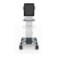
Mindray TE5 Operator's Manual (324 pages)
Diagnostic Ultrasound System
Brand:
Mindray
| Category:
Medical Equipment
| Size: 16.77 MB
Table of Contents
Table of Contents
3
Contents
7
Intellectual Property Statement
7
Responsibility on the Manufacturer Party
7
Warranty
8
Exemptions
8
Customer Service Department
8
Important Information
9
About this Manual
9
Notation Conventions
10
Operator's Manuals
10
Hardcopy Manuals
10
Software Interfaces in this Manual
10
Conventions
11
Product Differences
11
1 Safety Precautions
13
Safety Classifications
13
Meanings of Signal Words
14
Meaning of Safety Symbols
14
Safety Precautions
15
Latex Alert
22
Warning Labels
23
2 System Overview
25
Intended Use
25
Product Specifications
25
Imaging Mode
25
Environmental Conditions
26
Dimensions and Weight
26
System Configuration
26
Standard Configuration
26
Probes and Needle-Guided Brackets Available
27
Options
30
Peripherals Supported
31
Introduction of each Unit
32
Symbols
34
3 System Preparation
37
Move/Position the System
37
Power ON/OFF
38
Power the System on
38
Power the System off
39
To Power the System off Normally
39
To Shut down the System Directly if It Cannot be Done Normally
39
Standby
40
To Enter Standby
40
To Exit Standby
40
Under the Standby Status
40
Connecting the Power Cord
41
Powered by Batteries
41
Connecting a Probe
42
Disconnecting a Probe
43
Connecting the Footswitch
43
Connecting USB Devices
43
Installing a Graph/Text Printer
44
Connecting a Local Printer
44
Installing a Video Printer
46
Brightness and Contrast Adjustment
46
Display Position Adjustment
47
Basic Screen & Operation
48
Screen Display
48
Operating Panel
49
Ivocal
50
Vocal Command Inputting Procedures
50
4 Exam Preparation
55
Patient Information
55
To Enter the Patient Info Screen
55
To Exit the Patient Info Screen
55
New Patient Information
56
General Information
57
Exam Information
57
To Delete History Entered Information
57
Exam-Specific Information
58
Retrieve Patient Information
59
Retrieve from Istation
59
Functional Controls
59
Retrieve from Worklist
60
Select Exam Mode and Probe
61
Select the Imaging Mode
62
End an Exam
62
Activate an Exam
62
5 Image Optimization
63
Image Adjustment
63
Switching between Imaging Modes
63
Quickly Saving Image Settings
64
B Mode Image Optimization
66
B Mode Parameters
66
Basic Procedures for B Mode Imaging
66
Focus
68
Rotation/Invert (U/D Flip and L/R Flip)
68
Gray Map
69
Ibeam (Spatial Compound Imaging)
69
Itouch (Auto Image Optimization)
69
Tint Map
69
Dynamic Range
70
Iclear
70
Line Density
70
Persistence
70
Dual Live
71
Echo Boost
71
Middle Line
71
Patient Temperature
71
Basic Procedures for M Mode Imaging
72
M Mode Image Optimization
72
M Mode Image Parameters
72
Edge Enhance
73
M Soften
73
Speed
73
Basic Procedures for Color Mode Imaging
74
Color Mode Image Optimization
74
Color Mode Image Parameters
74
B/C Align
75
Color Gain
75
Frequency (Image Quality)
75
ROI Adjustment
75
Flow State
76
Packet Size
76
Smooth
76
Baseline
77
Color Map
77
Invert
77
Scale
77
Priority
78
Smart Tracking (ROI Auto Position/Steer in Color/Pw Mode)
78
Basic Procedures for Power Mode Imaging
79
Power Mode Image Optimization
79
Power Mode Image Parameters
79
Power Gain
80
Power Map
80
Basic Procedures for PW/CW Mode Exam
81
PW/CW Doppler Mode
81
PW/CW Mode Image Parameters
81
CW Focus Position
82
PW/CW Mode Image Optimization
82
Sv
82
Auto-Calculation
84
Basic Procedures
88
Contrast Imaging
88
Micro-Bubble Destruction
89
Parameter Area Display
89
Parameters
89
Timer
89
Image Saving
90
MIX Map
90
Contrast Imaging QA
91
Left Ventricular Opacification (LVO)
91
Measurement, Comments and Body Marks
91
Basic Operations of Contrast QA
92
Contrast QA Screen
92
QA Analysis Image Range
92
Setting ROI
92
Delete ROI
93
Ellipse ROI
93
Motion Tracking
93
Trace ROI
93
Curve Fitting
94
Adjustment of the M-Mark Line
95
Anatomical M Mode
95
Free Xros M Image Parameters
95
Free Xros M Mode
95
Display Format
96
Exit
96
Tdi
96
TDI Exam Protocol
96
TDI Image Optimization
97
TDI Image Parameters
97
Color M Mode
98
Enter Color M Mode
98
Exit Color M Mode
98
Image Parameters
98
3D Imaging
99
ROI and VOI
99
Curved VOI Adjustment
100
Mpr
100
ROI Size and Position
100
Basic Procedures for Smart 3D Imaging
101
Smart 3D
101
To Perform Smart 3D Imaging
101
Linear Scanning
103
Rocked Scanning
103
Smart 3D Acquisition Preparation
103
Activate MPR
104
Enter/Exit Image Viewing
104
Smart 3D Image Viewing
104
MPR Only
105
MPR Viewing
105
View Direction
106
Adjust VOI
107
Image Rendering Parameters
107
VOI off
107
VOI on
107
Axial Rotation
109
Comments and Body Marks
109
Image Zooming
109
Rotate an Image
109
6 Display & Cine Review
111
Splitting Display
111
Image Magnification
111
Izoom (Full Screen View)
111
Freeze/Unfreeze the Image
112
Imaging Mode Switching When Frozen
112
Cine Review
112
Entering/Exiting Cine Review
112
Manual Cine Review
113
Linked Cine Review
114
Image Compare
114
Auto Review
114
Reviewing All
114
Cine Saving
116
Preset
116
Setting Cine Length
116
Live Capture
116
7 Measurement
117
Basic Operations
117
To Enter/Exit Measurement
117
Measurement Results Window
117
General Measurements
118
Advanced Measurements
118
Measurement Accuracy
119
8 Physiological Signal
121
Ecg
122
ECG Operation Basic Procedures
122
ECG Triggering
122
Triggering Mode
123
ECG Review
123
Parameters Description
123
Review Principle
123
9 Annotations and Body Marks
125
Annotation Menu
125
Annotations Basic Procedures
125
Adding an Annotation Text
126
Adding Annotations
126
Moving Annotations
126
Typing Annotation Characters
126
Deleting Annotations
127
Modifying (Editing) Annotations
127
Modifying (Editing) Arrows
127
Modifying (Editing) Characters
127
Adding Voice Comments
128
Voice Comment Panel
128
Voice Comments
128
Adding Body Marks
129
Body Mark
129
Moving Body Marks
129
Voice Comment Review
129
Settings
130
10 Patient Data Management
131
Patient Information Management
131
Image File Management
131
Memory Media
131
Image Storage Preset
132
Quickly Saving Full Screen Images to the System
132
Set Image Size
132
Set Send/Print Image after Ending the Exam
132
Image Review and Analysis
133
Review an Image
133
Image Analysis
134
Ivision
134
Demonstration Item
135
Interval
135
Repetition
135
Exhibition
135
Sending Image Files
136
Report Management
137
Report Printing
138
Export Reports in Report Screen
138
Istation - Patient Data Management
139
Searching a Patient
140
Patient Data View & Management
140
Send Exam
141
Recycle bin
142
Istorage
142
Print
142
Image Printing
142
Back up Files Using the DVD Drive
143
Patient Task Management
144
Media Storage Task
144
Administration
145
Access Control
145
Enabling Access Control
145
System Login
145
Local Privilege Management
146
Adding a User/Assigning Privilege
146
To Change Users
146
Lock System
146
Modify Passwords
147
Configure Password Policy
147
LDAP Privilege Management
149
Adding a User
151
Deleting a User
151
Modifying the Member Name or Privileges
151
Logon Test
151
User Field Name
152
V-Access
153
Q-Path
153
Q-Path Basic Procedure
154
11 Dicom/Hl7
157
DICOM Preset
158
IP Preset
158
DICOM Local Preset
159
Server Setting Procedure
160
Service Preset
161
Storage Service Preset
161
Print Service Preset
163
DICOM Worklist Preset
165
MPPS Preset
165
Storage Commitment Preset
165
Query/Retrieve Preset
165
Verify Connectivity
166
Hl7Query Preset
166
DICOM Services
167
DICOM Storage
167
DICOM Print
168
DICOM Worklist
169
Mpps
169
Storage Commitment
169
Query/Retrieve
170
DICOM Media Storage
171
Data Restore
172
Structured Report
173
DICOM Task Management
173
12 Setup
175
System Preset
176
Region
178
Image Preset
181
Footswitch
182
POC Probe
183
Peripheral Preset
184
Image Settings
184
Maintenance
185
Exporting Setup Data
185
Other Settings
185
Security
186
System Information
195
Exam Related Preset
196
Exam Mode Preset
196
Comment Preset
197
Network Related Preset
198
Network Preset
199
Istorage Preset
199
Wireless Network Connection
200
Medtouch Preset
200
13 Probes and Biopsy
201
Probe Functions by Part
205
Operating Procedures
206
Orientation of the Ultrasound Image and the Probe Head
206
Wearing the Probe Sheath
209
Cleaning
212
Disinfecting with Sprays or Wipes
212
Probes Cleaning and Disinfection
212
Disinfecting by Immersion
213
Compatible Disinfectants
214
Glutaraldehyde-Based Sterilization Solution
214
Sterilization
214
Immerse the Probe in the Solution
215
Storage and Transportation
215
Biopsy Guide
216
Names of Parts
219
Needle-Guided Brackets
219
Ngb-007
220
Plastic/Needle Detachable Needle-Guided Bracket
220
Basic Procedures for Biopsy Guiding
227
Inspection of the Needle-Guided Bracket
228
Installing the Needle-Guided Bracket
228
Needle-Guided Bracket Inspection and Installation
228
Metal/Needle Detachable Needle-Guided Bracket
229
Plastic Needle-Guided Bracket
229
Biopsy Menu
236
B/Ineedle
237
Ineedle (Needle Visualization Enhancement)
237
Needle Direction
237
To Enter/Exit Ineedle
237
Adjusting the Needle Mark Is Necessary before each Biopsy Procedure
238
Verifying the Biopsy Guide Line
238
Removing the Needle-Guided Bracket
239
Clean and Sterilize the Needle-Guided Bracket
243
Disposal
244
Middle Line
244
Espacial Navi
245
In Plane Needle Guidance GUI
246
Interface
246
Out of Plane Needle Guidance GUI
247
Es.navi Settings Menu
248
Preparation before Needle Guidance
248
Procedure
249
14 DVR Recording
251
Sending Image
251
DVR Video Replaying
251
Replay on the Ultrasound System
252
15 Acoustic Output
253
Concerns with Bioeffects
253
Prudent Use Statement
253
ALARA Principle (as Low as Reasonably Achievable)
253
MI/TI Explanation
254
Basic Knowledge of MI and TI
254
Mechanical Bioeffect and Thermal Bioeffect
254
MI (Mechanical Index)
254
Acoustic Power Setting
255
MI/TI Display
255
Acoustic Power Adjustment
255
Default Acoustic Power Setting
255
Acoustic Power Control
256
Derated Ultrasonic Output Parameters
256
Direct Controls
256
Indirect Controls
256
Measurement Uncertainty
257
Limits of Acoustic Output
257
Differences between Actual and Displayed MI and TI
257
References for Acoustic Power and Safety
258
Advertisement
Mindray TE5 Operator's Manual (312 pages)
Brand:
Mindray
| Category:
Speaker System
| Size: 16.37 MB
Table of Contents
Intellectual Property Statement
3
Responsibility on the Manufacturer Party
3
Warranty
4
Exemptions
4
Customer Service Department
4
Important Information
4
About this Manual
5
Meaning of Signal Words
5
Hardcopy Manuals
6
Software Interfaces in this Manual
6
Conventions
6
Operator's Manuals
6
Table of Contents
7
Important Information
15
Safety Precautions
15
Latex Alert
21
Parts that Can be Used Within Patient Environment
21
System Overview
23
Intended Use
23
Contraindication
23
Safety Classifications
23
Product Specifications
24
Power Supply
24
Environmental Conditions
24
Dimensions and Weight
25
Product Differences
25
System Configuration
25
Standard Configuration
25
Probes and Needle-Guided Brackets Available
26
Options
27
Peripherals Supported
29
CIVCO Probe Sheath and Brackets
29
Introduction of each Unit
30
ECG Module
32
Basic Operations
32
Screen Display
32
Control Panel
34
Select Exam Mode and Probe
35
Select the Imaging Mode
35
Imaging Adjustment
36
Quickly Saving Image Settings
36
Annotations and Body Marks
37
Ivocal
38
Splitting Display
40
Image Magnification
40
Izoom (Full Screen View)
41
Freeze/Unfreeze the Image
41
Imaging Mode Switching When Frozen
42
Cine Review
42
Entering/Exiting Cine Review
42
Manual Cine Review
43
Auto Review
44
Linked Cine Review
44
Symbols and Warning Labels
44
System Preparation
47
Move/Position the System
47
Connecting the Power Cord
47
Connecting Power
47
Powered by Batteries
47
Power ON/OFF
48
Check before Powering on
49
Power the System on
49
Check the System after It Is Powered on
50
Power the System off
51
Standby
51
Monitor Brightness/Contrast Adjustment
52
Display Position Adjustment
52
Connecting/Disconnecting a Probe
52
Connecting a Probe
53
Disconnecting a Probe
54
Connecting USB Devices
54
Connecting the Footswitch
55
Installing a Printer
55
Connecting a Graph/Text Printer
55
Connecting a Video Printer
55
Setup
57
System Preset
57
Region
57
General
58
Image Preset
59
Measure
60
Footswitch Function
61
Probe Function
61
Access Control
62
Scan Code Preset
64
Peripheral Preset
67
Maintenance
67
Ivision
69
Security
69
System Information
71
Exam Related Preset
71
Exam Mode Preset
71
Measurement Preset
71
Body Mark/Comment Preset
73
Iworks Preset
73
Network Related Preset
74
DICOM Local Preset
74
DICOM Service Preset
76
Egateway Preset
81
Network Preset
83
Q-Path Preset
83
Exam Preparation
85
Patient Information
85
New Patient Information
85
Retrieve Patient Information
86
Activate an Exam
86
End an Exam
86
Image Acquisition
87
B Mode
87
B-Mode Image Scanning
87
B-Mode Image Parameters
87
Color Mode
90
Color Mode Image Scanning
90
Color Mode Image Parameters
90
Power Mode
92
Power Mode Image Scanning
92
Power Mode Image Parameters
92
M Mode
93
M Mode Image Scanning
93
M Mode Image Parameters
93
Color M Mode (CM)
94
CM Image Scanning
94
CM Image Parameters
94
Anatomical M Mode
94
Linear Anatomical M (Free Xros M)
95
Anatomical M Mode Parameters
95
PW/CW Mode
95
PW/CW Mode Image Scanning
95
PW/CW Mode Image Parameter
96
Tdi
97
TDI Mode Image Scanning
98
TDI Mode Image Parameters
98
Iscape View (Real-Time Panoramic Imaging)
98
Basic Procedures for Iscape Imaging
99
Image Review
100
Evaluate Image Quality
100
Cine Review
101
Smart B-Line
101
Basic Procedures for Smart B-Line
101
Overview
102
Smart VTI
103
Smart IVC
104
Contrast Imaging
105
Basic Procedures for Contrast Imaging
106
Left Ventricular Opacification
106
Image Parameters
107
Image Saving
108
Micro Flow Enhancement
108
Contrast Imaging QA
109
Auto GA
112
Smart FHR OB1
112
Iworks (Auto Workflow Protocol)
113
Overview
113
Normal Iworks Basic Procedure
113
View Operation
114
Manual Examination
114
Insert
115
Smart 3D
117
Overview
117
Terms
117
ROI and VOI
118
Render Mode
118
Mpr
119
Note before Use
120
Smart 3D Image Acquisition
122
Smart 3D Image Viewing
123
Image Saving
127
Physiological Unit Signal
129
Ecg
129
ECG Review
131
Review Principle
131
Linked Review of Waveforms, M/D Images and 2D Images
131
Parameter Description
131
Measurement, Annotations and Body Mark
133
Measurement
133
Annotations
135
Adding Annotations
135
Moving Annotations
135
Modifying (Editing) Annotations
136
Deleting Annotations
136
Body Mark
136
Adding Body Mark
137
Moving Body Marks
137
Deleting Body Marks
137
Voice Comments
137
Voice Comment Panel
137
Adding Voice Comments
138
Voice Comment Review
138
Patient Data Management
139
Image File Management
139
Storage Media
139
Image File Formats
139
Image Storage Setting
140
Saving Full Screen Images to the System
140
Image Review and Analysis
140
Sending Image Files
141
Report Management
141
Report Storage
141
Importing, Exporting and Sending a Report
141
Istation - Patient Data Management
142
Searching a Patient
142
Patient Data View & Management
142
Image Compare
143
Recycle bin
143
Istorage
144
Print
144
Image Printing
144
Report Printing
145
Back up Files Using the DVD Drive
145
Patient Task Management
145
V-Access
147
Q-Path
147
Dicom/Hl7
149
DICOM Storage
149
Send Images on Istation/Review
149
To Send Images to Storage after an Exam Ends
150
DICOM Print
150
Print Images on Istation/Review
150
To Send Images to DICOM Print after an Exam Ends
150
To Print Images after Saving Image
150
Worklist
150
Mpps
151
Storage Commitment
151
After Sending Images on the Istation Screen
151
Storage Commitment after an Exam Ends
152
Query/Retrieve
152
DICOM Media Storage
152
Media Storage
152
Media Review
153
Data Restore
153
Showcase Recording
153
Structured Report
153
Send Structured Reports for Storage
154
Back up Structured Report
154
Probes and Biopsy
155
Probes
155
Laparoscopic Probe
158
Probe Functions by Part
160
Orientation of the Ultrasound Image and the Probe Head
161
Procedures for Operating
162
Wearing the Probe Sheath
164
Probes Cleaning and Disinfection/Sterilization
166
Cleaning the Probe Cable and Connector
171
Probe Environmental Conditions
171
Storage and Transportation
173
Biopsy Guide
174
Needle-Guided Brackets Available
176
Needle-Guided Bracket Inspection and Installation
176
Verifying the Biopsy Guide Line
204
Starting the Biopsy Procedure
205
Clean and Sterilize the Needle-Guided Bracket
206
Storage and Transportation
208
Disposal
208
Middle Line
208
Espacial Navi
209
Interface
209
Preset
211
Preparation before Needle Guidance
211
Procedure
212
DVR Recording
213
Start Recording
213
Sending Image
213
DVR Video Replaying
214
Replay on PC
214
Replay on the Ultrasound System
214
System Maintenance
215
Daily Maintenance
215
Cleaning the System
215
Cleaning the Peripherals
218
Disinfecting the Main Unit
218
Checking the Probe
222
Checking the Power Cable and Plug
222
Checking Appearance
223
System Hard Drive Backup
223
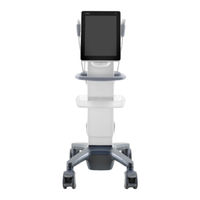
Mindray TE5 Operator's Manual (337 pages)
Diagnostic Ultrasound System
Brand:
Mindray
| Category:
Medical Equipment
| Size: 6.77 MB
Table of Contents
Table of Contents
3
Contents
7
Intellectual Property Statement
7
Responsibility on the Manufacturer Party
7
Warranty
8
Exemptions
8
Customer Service Department
8
Important Information
9
About this Manual
10
Notation Conventions
10
Operator's Manuals
10
Hardcopy Manuals
10
Software Interfaces in this Manual
10
Conventions
11
Product Differences
11
Safety Precautions
13
Safety Classifications
13
Meanings of Signal Words
14
Meaning of Safety Symbols
14
Safety Precautions
15
Latex Alert
21
Warning Labels
23
System Overview
25
Intended Use
25
Contraindication
25
Product Specifications
25
System Configuration
26
Introduction of each Unit
33
Symbols
35
System Preparation
37
Move/Position the System
37
Power ON/OFF
38
Connecting the Power Cord
41
Connecting a Probe
42
Connecting the Footswitch
43
Connecting USB Devices
43
Installing a Graph/Text Printer
44
Installing a Video Printer
45
Brightness and Contrast Adjustment
45
Display Position Adjustment
46
Basic Screen & Operation
47
Exam Preparation
53
Patient Information
53
Select Exam Mode and Probe
60
Select the Imaging Mode
61
End an Exam
61
Activate an Exam
61
Image Optimization
63
Imaging Mode
63
B Mode Image Optimization
66
M Mode Image Optimization
72
Color Mode Image Optimization
75
Power Mode Image Optimization
80
PW/CW Doppler Mode
82
Smart B-Line
88
Smart VTI
90
Smart IVC
91
Contrast Imaging
94
Anatomical M Mode
101
Iscape View (Real-Time Panoramic Imaging)
102
Tdi
106
Color M Mode
108
Imaging
109
Display & Cine Review
121
Splitting Display
121
Image Magnification
121
Izoom (Full Screen View)
121
Freeze/Unfreeze the Image
122
Cine Review
122
Image Compare
124
Cine Saving
125
Preset
125
Measurement
127
Basic Operations
127
General Measurements
128
Advanced Measurements
129
Measurement Accuracy
130
Physiological Signal
131
Ecg
132
Parameters Description
133
Annotations and Body Marks
135
Annotations
135
Voice Comments
137
Body Mark
139
Settings
139
Patient Data Management
141
Patient Information Management
141
Image File Management
141
Report Management
147
Istation - Patient Data Management
149
Recycle bin
152
Istorage
152
Print
152
Back up Files Using the DVD Drive
153
Patient Task Management
154
Administration
155
V-Access
162
Q-Path
163
Dicom/Hl7
167
DICOM Preset
168
Verify Connectivity
178
DICOM Services
180
DICOM Media Storage
184
Structured Report
186
DICOM Task Management
186
Setup
187
System Preset
188
Exam Related Preset
201
Network Related Preset
204
Probes and Biopsy
209
Probes
209
Biopsy Guide
226
Middle Line
260
Espacial Navi
260
DVR Recording
267
Advertisement
Mindray TE5 Operator's Manual (103 pages)
Diagnostic Ultrasound System
Brand:
Mindray
| Category:
Medical Equipment
| Size: 1.58 MB
Table of Contents
Table of Contents
3
Intellectual Property Statement
7
Preface
8
Safety Precautions
9
1 Overview
11
Basic Operations and Buttons
11
Measurement Menu
11
Measurement Location
12
Measurement Tool
12
Measurement, Calculation and Study
13
Measurement Caliper
14
Results Window
14
Results Display
14
Moving the Results Window
15
Cross-Window Measurement
15
Report
15
Viewing Reports
16
Editing Reports
17
Printing Reports
18
2 Measurement Preset
19
Basic Preset Procedures
19
Measurement Parameters Preset
19
Obstetric Preset
20
Obstetric Formula
20
Obstetric Preset Operations
24
Measurement Preset
25
General Basic Measurement Preset
25
Advanced Measurement Preset
27
Setting Default Report Template
28
3 Basic Measurement
29
Basic Measurement Procedures
29
Basic Measurement Tools
29
Depth
29
Distance
29
Angle
30
Area & Circumference
30
Volume
31
Ratio (D)
31
Ratio (A)
31
Time
32
Slope
32
Velocity
33
D Trace
33
D Trace (Car)
36
Ps/Ed
36
Volume Flow
36
Imt
37
References
38
4 Abdomen
39
Abdomen Exam Preparations
39
Basic Abdomen Measurement Procedures
39
Abdomen Measurement Tools
40
Abdomen Measurement Operations
42
Abdomen Exam Report
42
5 Obstetrics
43
Obstetric Exam Preparations
43
Basic Measurement Procedures
43
Clinical GA
43
Ultrasound GA
44
Obstetric Measurement Tools
45
Obstetric Measurement Operations
46
Measurement Tool Operations
47
Calculation Tool Operations
47
Study Tool Operations
47
Obstetric Exam Report
47
Fetal Biophysical Profile
48
References
48
6 Cardiology
57
Cardiac Exam Preparations
57
Basic Cardiac Measurement Procedures
57
Cardiac Measurement Tools
58
Cardiac Measurement Operations
61
Measurement Tool Operations
61
Calculation Tool Operations
62
Study Tool Operations
62
Cardiac Exam Report
72
References
72
7 Vascular
75
Vascular Exam Preparations
75
Basic Vascular Measurement Procedures
75
Vascular Measurement Tools
75
Vascular Measurement Operations
77
Measurement Tool Operations
77
Calculation Tool Operations
77
Study Tool Operations
78
Vascular Exam Report
79
References
79
8 Gynecology
81
Gynecology Exam Preparations
81
Basic Gynecology Measurement Procedures
81
Gynecology Measurement Tools
81
Gynecology Measurement Operations
82
Measurement Tool Operations
82
Calculation Tool Operations
82
Study Tool Operations
83
Gynecology Exam Report
84
References
84
9 Urology
85
Urology Exam Preparations
85
Basic Urology Measurement Procedures
85
Urology Measurement Tools
85
Urology Measurement Operations
86
Measurement Tool Operations
87
Calculation Tool Operations
87
Study Tool Operations
88
Urology Exam Report
89
References
89
Advertisement
Related Products
Mindray Panorama TELEPACK-608 SpO2
Mindray TE7
Mindray TE Air
Mindray TE7T
Mindray TE7S
Mindray TE7 Pro
Mindray TE5T
Mindray TE5S
Mindray TE5 Pro
Mindray TM70
Mindray Categories
Medical Equipment
Diagnostic Equipment
Measuring Instruments
Monitor
Water Pump
More Mindray Manuals
Login
Sign In
OR
Sign in with Facebook
Sign in with Google
Upload manual
Upload from disk
Upload from URL