
Subscribe to Our Youtube Channel
Related Manuals for Leica DMI6000B


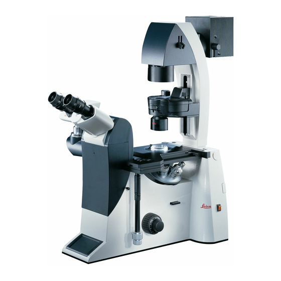
Summary of Contents for Leica DMI6000B
- Page 1 Leica DMI3000B Leica DMI4000B Leica DMI6000B Operating Manual Bedienungsanleitung M I C R O S Y S T E M S...
- Page 2 Published May 2005 by/ Herausgegeben Mai 2005 von: Leica Microsystems Wetzlar GmbH Ernst-Leitz-Straße D-35578 Wetzlar (Germany) Responsible for contents/ Verantwortlich für den Inhalt: Bernard Kleine (Marketing CMS, Life Science Research Microscopy, Product Management) Holger Grasse (Safety Officer according to MPG §30/ Sicherheitsbeauftragter nach MPG §30)
- Page 3 Leica DMI3000B Leica DMI4000B Leica DMI6000B Operating Manual...
- Page 4 (in whole or in part) by print, photocopy, microfilm or other method (in- cluding electronic systems) is not allowed with- out express written permission from Leica Microsystems Wetzlar GmbH. The term “Windows” may appear in the follow- ing text without further identification.
-
Page 5: Table Of Contents
3.1 General Safety Notes ....... 6.14 Inserting the Front Module Slider ..3.2 Electrical Safety ........10 6.15 Installation of the Polarizer Overview of the Leica DMI6000 ..... 12 and Analyzer ..........50 6.16 Optional Accessories ....... 52 4.1 Specifications ..........12 6.17 Connection to the... - Page 6 Contents Operation ............ 74 Troubleshooting ......... 91 8.1 Switching On ..........74 8.2. Contrast Methods ........76 10. Care of the Microscope ......95 8.2.1 Bright Field (TL) ....... 76 10.1 Dust Cover ..........95 8.2.2 Phase Contrast (TL) ......77 10.2 Cleaning ............
-
Page 7: Important Notes About This Manual
It must therefore be kept safely for bled, put into operation or used. future reference. A separate manual is available on CD-ROM cov- ering the operation of the Leica Application Suite (LAS). Text symbols, pictograms and their meanings: (1.2) Numbers in parentheses, such as “(1.2)”, corre-... -
Page 8: Intended Purpose Of The Microscope
Caution! The Leica DMI series is a further development of Leica’s proven inverted research microscopes.- The manufacturer assumes no liability for It is designed for cellular and tissue examina-... -
Page 9: Safety Notes
EN 61010-1:2001, safety and potential hazards. IEC 1010-1:2001, The responsible Leica affiliate or the main Safety regulations for electrical measuring, con- plant in Wetzlar must be consulted when- trol, and laboratory devices. ever the device is altered, modified or used in conjunction with non-Leica components that are outside of the scope of this manual. -
Page 10: Electrical Safety
Power input: see CTRxxxx tive conductor potential by connecting them Fuses: see CTRxxxx to the ground screw on the back of the Leica Ambient temperature: 15–35 C CTRxxxx electronics boxes. For connections Relative humidity: max. 80% to 30 C... - Page 11 3. Safety Notes Caution! The microscope’s electrical accessory com- ponents are not protected against water. Water can cause electric shock. Caution! Protect the microscope from excessive tem- perature fluctuations. Such fluctuations can lead to the accumulation of condensation, which can damage the electrical and optical components.
-
Page 12: Specifications
Fluo/DIC, Fluo/PH Leica DMI Series Transmitted Light Axis For the Leica DMI3000 B, a manual version of this illumination arm is always a component of the stand. • Manual and coded transmitted-light illumination arm with inte- grated mechanical tilt mechanism to provide adequate space... - Page 13 4. Overview of the Instrument Incident light axis Leica DMI4000 B and Leica DMI6000 B • automatic Illumination Manager (aperture, field diaphragm, intensity, process switching) • motorized shutter (switching speed < 50 ms) • lamp housing mount for up to 3 interchangeable light sources •...
- Page 14 4. Overview of the Instrument Objective Turret Leica DMI4000 B • manual and coded • 6x for objectives with M25 thread and 45mm parfocal distance • for DIC: motorized or manual/coded Wollaston prism carousel Leica DMI3000 B • manual • 6x for objectives with M25 thread and 45 mm parfocal distance •...
- Page 15 4. Overview of the Instrument Condensers Leica DMI4000 B and Leica DMI6000 B (identical for Leica DMI3000 B, but manual) • motorized and coded or manual and coded • motorized or manual aperture diaphragm • contrast methods: BF, DF, PH, DIC, Pol, IMC, IPH •...
- Page 16 4. Overview of the Instrument Observation ports Leica DMI3000 B (a manual side port is a standard feature of the Leica DMI3000 B stand) • manual • left side port (80% transmission) Controls Leica DMI4000 B and Leica DMI6000 B •...
- Page 17 4. Overview of the Instrument Interfaces Leica DMI4000 B and Leica DMI6000 B • 2 x RS232C • 2 x USB • 4 x external/internal peripherals • CTR boxes • SmartMove Software tools Leica DMI4000 B and Leica DMI6000 B •...
- Page 18 4. Overview of the Instrument Fig. 1 Left side, Leica DMI4000 B and DMI6000 B Eyepiece 11 Variable function buttons Eyepiece tube 12 Left side port Top port 13 Booster lens (fluorescence microscopes only) Intermediate pupil interface 14 Lamp mount (fluorescence microscopes only)
- Page 19 4. Overview of the Instrument Fig. 2 Right side Leica DMI4000 B and DMI6000 B E-Focus buttons (Leica DMI6000 B only) Analyzer slot Focus wheel (motorized Leica DMI6000 B, Centering window (fluorescence microscopes only) manual (fine) Leica DMI4000 B) Field diaphragm centering...
- Page 20 4. Overview of the Instrument Fig. 3 Front view Leica DMI4000 B and Leica DMI6000 B LeicaScreen Front control panel Port switching Top port Manual transmitted-light filters Bertrand lens centering...
- Page 21 Shutter Travel in y 100% light to all eyepieces Focus Port selection Variable function buttons Magnification selection (preassigned at factory) 1x tube lens Fig. 4 General view Leica DMI4000 B and Leica DMI6000 B with SmartMove remote control module...
- Page 22 4. Overview of the Instrument Fig. 5 Leica DMI3000 B left view Eyepiece Eyepiece tube Top port Intermediate pupil interface Light intensity Focus wheel Left side port DIC objective prism disk Condenser head 10 Condenser base 11 Field diaphragm 12 Transmitted-light lamp housing...
- Page 23 4. Overview of the Instrument Fig. 6 Leica DMI3000 B right view Focus wheel Analyzer slot Objective turret On/Off switch Stage with attachable mechanical stage...
- Page 24 4. Overview of the Instrument Fig. 6 Leica DMI3000 B front view Port switching + Bertrand lens Top port Manual transmitted-light filters Bertrand lens centering...
- Page 25 4. Overview of the Instrument Fig. 6b Leica DMI3000 B Manual magnification changer Bertrand lens centering Top port Manual transmitted-light filters Interchangeable transmitted-light lamp housing On/Off switch...
-
Page 26: Unpacking The Microscope
• Illumination arm • Assembly tools • Specimen stage • Additional accessories such as filter cubes, • CD with Leica Application Suite (LAS) soft- etc. depending on feature set ware package • Instructions and list of microscope presets The Leica CTRxxxx electronics box, the... - Page 27 5. Unpacking the Microscope Please carefully compare the contents of the de- livery to the packing slip, delivery note or in- Caution! voice. We urgently recommend storing a copy of these documents with the manual to ensure that Do not connect the microscope or periph- you have information on the time and scope of erals to an AC power source at this time delivery handy for subsequent orders or service...
- Page 28 5. Unpacking the Microscope Transport For shipping or transporting the microscope and its accessory components, the original packaging should be used. As a precaution to prevent damage from vibra- tions, the following components should be dis- assembled and packaged separately: •...
-
Page 29: Assembling The Microscope
The microscope components* are logically as- sembled in this order: If possible, the microscope should be assem- bled and set up with the assistance of Leica • Transmitted-light illumination carrier sales or service personnel. • DIC module and DIC objective prisms A small number of universal screwdrivers which •... -
Page 30: Installation Of The Transmitted-Light Illumination Carrier
6. Assembly 6.2 Installation of the Leica DMI4000 B and Leica DMI6000 B transmitted-light illumination carrier (DL) Connect the electronics cable to one of the sockets, EXT1 - EXT4. Wipe the installation surface on the microscope (8.3) with a dry cloth. Tip the illumination carrier The transmitted-light lamp housing for 12 V (8.1) back slightly and install it so that the pin... -
Page 31: Installation Of The Dic Module And Dic Objective Prisms
Allen screws. please continue with Chapter 6.4. Note: insert the prism disk with the electron- In the Leica DMI series microscopes, the DIC ics board facing down. Do not touch the elec- prisms are already installed in the DIC disk be- tronics (especially the contacts) with your low the objective turret (Fig. - Page 32 6. Assembly 6.4 Installation of Specimen Stages Fig. 14 Mechanical 3-plate stage A wide range of specimen stages are available. The most important are the following: • Fixed stage (248 mm x 204 mm) (Fig. 13): normal, heating and temperature-controlled, with and without attachable mechanical stage •...
- Page 33 6. Assembly The assembly of these stages is identical. The • Use a clean cloth to remove dust and packing stages are solidly attached to the microscope by material residue from the stand’s contact sur- three screws. In the case of fixed stages, an at- face for the stage.
- Page 34 Leica’s technical service. Fixed stage Next, remove one or more of the ordered insert Attachable mechanical stages designed to ac- frames (Fig.
- Page 35 6. Assembly Manual fixed micromanipulation stage To install the attachable mechanical stage for the manual fixed micromanipulation stage (Fig. 24), proceed as you would for the attach- able mechanical stage of the standard stage. Fig. 24 Installation of attachable mechanical stage The insert frames (Fig.
- Page 36 6. Assembly Motorized 3-plate or scanning stages Caution: 3-plate stages and scanning stages: after in- Press the spring clip into place only from the stalling the stage, connect the included stage side. cable (for motorized stages) first to the socket on the stage, then to the CRT6000 or CTR6500 Do not press the insert onto the spring clips di- box.
- Page 37 6. Assembly Rotating Stage and Insert Frames for Coverslips Fig. 30 Rotating stage Attachable mechanical stage The rotating stage (Fig. 30) is also mounted with Screws for stage mounting 3 screws (30.2). Rotate the stage to make all of Washers the threaded holes accessible.
- Page 38 6.5 Installation of Condensers the adjusting key (39.2) into the left-hand centering screw of the disk. It may protrude a All condensers of the Leica DMI series are maximum of 1 mm into the opening. equipped with a 7-position turret disk that can...
- Page 39 Press the cheeks of the tool to grasp the light ring (Fig. 39a). • Note the number of the opening and the light ring designation for entry into the Leica Appli- • Two guide hooks are located on the underside cation Suite (LAS).
- Page 40 6. Assembly Please continue reading if you also have to in- • Grasp the prism to be installed with the con- stall IC prisms. Otherwise, skip to the next sec- denser tool (the lettering must face upward tion. and be legible) so that the tab of the prism ring is positioned to the center of the tool’s cam and the upper edge of the prism is lying flat in Installation of IC prisms...
-
Page 41: Installation Of Condensers
• Note the number of the opening and the prism designation for entry into the Leica Applica- tion Suite (LAS). • Remove the adjusting key and close the con- denser. - Page 42 6. Assembly Condenser heads Four different condenser heads are available: S1/1.40 oil S1/0.90 dry S23/0.53 S28/0.55 Condenser heads 3 and 4 are screwed directly into the condenser body. A spacer ring (42.2) must be screwed into the thread at the bottom of the condenser body prior to installing condenser heads 1 and 2.
-
Page 43: Installation Of Eyepieces
Note: For details on the exact positions of the objec- tives, please refer to the enclosed identifica- We recommend running a teach-in via the Leica tion sheet. Application Suite (LAS) software when using eyepieces not included in the scope of delivery. -
Page 44: Installation Of Filters In The Illumination Arm
6.8 Installation of filters in the illumination arm 6.9 Installing the transmitted-light lamp housing The Leica DMI series is equipped with a filter magazine to accommodate two 40 mm dia. filters • Place the lamp housing in the transmitted light as a standard feature. -
Page 45: Installation And Replacement Of The ...... Transmitted-Light Lamps
6. Assembly 6.10 Installation and replacement of the • Lift the housing off (Fig. 50b). transmitted-light lamps: 107 or 107/2 lamp housing • Remove the lamp. This lamp housing is used with a 12V 100W halo- Caution! gen lamp, which is already mounted. In case the lamp has to be removed: Do not remove the new lamp’s dust cover until you have installed the lamp. - Page 46 The lamp housing or coupling is then mounted on the connector, which is also held by four screws. Fig. 52 Rear view, Leica DMI4000 B and DMI6000 B Fig. 54 Lamp housing 106z Installation point for lamp housing mount...
- Page 47 6. Assembly If a booster lens is included in the scope of de- livery, insert it into the rear stand opening at the left or right, depending on the stand model. The booster slide has several positions: 1. Slide pulled out: no effect 2.
-
Page 48: Lamp Housing 107 Or 107/2
This lamp housing is suitable for use with a 12 V 100 W halogen lamp or a variety of gas discharge lamps. Caution! Caution! Leica DMI4000 B and Leica DMI6000 B: Light sources pose a potential irradiation Make sure to follow the instructions and risk (glare, UV-radiation, IR-radiation). - Page 49 6. Assembly Inserting gas discharge lamps (Hg and Xe) in the 106z lamp housing Hg and Xe lamps are powered by separate sup- ply units. Please also read the separate instruction manual provided with these supply units. The following gas discharge lamps may be used and require different supply units and lamp mounts (Fig.
- Page 50 6. Assembly Caution! Caution! Make sure to follow the safety notes on Do not remove the burner’s dust cover until you page 48. have installed the lamp. Avoid fingerprints on the lamp. Sweat from your fingers on the glass • To open the 106 z lamp housing, unscrew the will shorten the life of the lamp significantly.
- Page 51 6. Assembly • Insert the lamp mount, with the burner in- stalled, into the lamp housing and tighten it with the screws (63.8). • Test the adjustment of the collector (63.2): Do not touch the power supply while perform- ing these actions. When closing the lamp housing, ensure that the pins of the contact plug engage in their sockets (63.9).
-
Page 52: Equipping The Incident Light Turret Disk
Opening the fluorescence drawer ginning with the assembly of the turret disk. 1 Analyzer slot Leica DMI4000 B and Leica DMI6000 B: The fluorescence drawer is located on the right side of the stand. Before opening this drawer, remove the cap below the drawer covering the analyzer slot (66.1). - Page 53 6. Assembly The positions in the turret disk are numbered. • For the next cube, close the drawer to the Depending on your equipment, the individual fil- point that the disk is once again free to turn. ter and reflector cubes have already been as- Once you have reached the next position, signed to specific positions at the factory.
-
Page 54: Inserting The Front Module Slider
6. Assembly 6.14 Inserting the Front Module Slider 6.15 Installation of the polarizer and analyzer If your microscope is prepared for integrated Installed at the factory. modulation contrast or integrated phase con- To change the components, proceed as follows: trast, a front module (possibly in conjunction with a manual magnification changer) will be in- Motorized condenser: tegrated in the stand. - Page 55 6. Assembly Analyzer for incident light and transmitted light. • Remove the cap (Fig. 72) on the right side of the stand (under the fluorescence drawer). • Insert the analyzer into the receptacle until it latches in place (Fig. 73.1). Fig.
-
Page 56: Optional Accessories
Fig. 75 C-mount 0.63x Note: When using a C-mount or Vario mount, run a teach-in via the Leica Application Suite (LAS) software. Connecting multiple cameras Two or more cameras – for example a digital and an analog camera – can be adapted as required. -
Page 57: Connection To The Electronics Box Ctr6000
6.17 Connection to the Electronics Box CTR 4000 electronics box CTR4000, CTR6000 or CTR6500 The Leica DMI 4000 B is supplied with the CTR4000 electronics box. The power supply for The Leica DMI 3000 B is supplied without an the microscope is located in this box. Two sock- electronics box. -
Page 58: Connection To The Computer
Note: Note: These electronics boxes must not be used with To start the Leica Application Suite (LAS), en- other stands. The serial number of the associated sure that the COM1 serial port is not in use by stand has been recorded on the back of the elec- another program or driver. -
Page 59: Startup
7. Start-up 7. Start-up 7.1 Functional Principle (Leica DMI4000 B and Leica DMI6000 B) Thanks to its intelligent automation, the Leica DMI4000B and DMI6000B can be controlled using a variety of control elements. 1. Intelligent automation • Switching between contrast methods at the touch of a button. Light rings, DIC prisms, etc. - Page 60 7. Start-up The table on the following page provides an overview of the microscope functions and their Note: (reset function) controls. The microscope can be reset to its factory de- fault programming: • With the stand switched off, press the top three variable function buttons on the left side of the stand.
- Page 61 7. Start-up Function Fixed Variable SmartMove Software (DMI4000 and DMI6000 B) Function Function Function Rotary buttons buttons buttons knobs Stand Stand 4000 6000 4000 6000 4000 6000 4000 6000 4000/6000 Select contrast method Change transmitted light/incident-light axis Change to objective Teach-in parfocality Change operating mode (dry/imm) Illumination Manager...
- Page 62 7. Start-up Possible assignments for variable function buttons on stand and SmartMove For Leica DMI4000 B and Leica DMI6000 B: Function button Function Bright field transmitted light Phase contrast transmitted light Interference contrast, transmitted light Dark field transmitted light Integrated modulation contrast...
-
Page 63: Switching On
Off switch . The signal lamp is lit when the in- light and phase rings have been pre-centered at strument is ready. (For the Leica DMI3000 B the factory. It may be necessary to correct the please continue at 7.4. Function Buttons on... - Page 64 7. Start-up 7.3 The LeicaDisplay Pictograms (Leica DMI 4000 B and DMI 6000 B) The screen displays the microscope’s current Contrasting method settings. The content of the display depends on the features of the individual microscope. Objective/ For information on the abbreviations used, please...
-
Page 65: The Function Buttons On The Stand
(a version of the Leica DMI3000 B with mir- rored focus controls is also available) • Light intensity: the transmitted-light intensity... - Page 66 For information on the abbreviations used, please refer to the list p. 62. Note: The Leica Application Suite (LAS) software is required for changing the button assignments. Possible functions*: CHANGE CUBE CW CHANGE CUBE CCW...
- Page 67 Switching to the bottom port: the buttons again or wait 3 seconds. via the variable function buttons (Leica DMI6000 B only), switching to top port: manually. Focus buttons (Fig. 88) (DMI6000 B only) SHUTTER Opens and closes the shutter (87.3).
-
Page 68: The Smartmove Remote Control Module
Use the knobs 89.1 and 89.2 to move the stage in • Focus on the specimen using the focus wheels. X and Y directions. The image is focused using the knob 89.3 (Leica • Adjust the light intensity. DMI6000 B only). - Page 69 • Place a specimen on the stage. All required functions can be executed at the touch of a button with the Leica DMI6000 elec- • Focus the specimen using the SmartMove or tronic microscope. (See Chapter 8, Operation).
- Page 70 7. Start-up • Switch to the 10x objective (if not present, the • You should see light when looking through the 20x objective). eyepieces at this point. • Ensure that the condenser is at the correct If the light is too bright, reduce it as required. height.
- Page 71 7. Start-up field of view. The black edges of the image To see the aperture diaphragm, remove an should have the same distance to the outer eyepiece tube and look directly into the tube. edge of the field of view on all sides. If not, Your eye should be around 10 to 20 cm from recenter the image with the centering screws.
-
Page 72: Incident Light - Fluorescence
7. Start-up 7.6.2 Incident Light - Fluorescence Adjusting the field diaphragm (Leica DMI4000 B and Leica DMI6000 B) • Close the field diaphragm with the FD button (84.4) or manually until the edge of the dia- Suitable aperture and field diaphragm values phragm (round or rectangular) appears in the have been preset for each objective. -
Page 73: Checking Phase Contrast Rings
7. Start-up 7.7 Checking Phase Contrast Rings Leica DMI4000 B and Leica DMI6000 B: • Press the BF (bright field) button (one of the If your microscope is equipped for phase con- variable function buttons on the stand). trast, light rings to match your objectives will be installed in the condenser. - Page 74 Leica DMI3000 B: • Select the light ring for the active objective on • Remove the centering keys after centering. the condenser. Leica DMI4000B and Leica DMI6000B: Fig. 96 Condenser with motorized polarizer • Press the PH (phase contrast) button (one of...
- Page 75 7. Start-up Integrated phase contrast with bright field Leica DMI4000 B and Leica DMI6000 B: objectives via front slider • Press the PH (phase contrast) button (one of the variable function buttons behind the focus When choosing an objective suitable for phase wheels).
-
Page 76: Setting The Motorized Polarizer
• Turn the polarizer until you have the optimal dark position. Note: Leica DMI4000 B and Leica DMI6000 B: For manual condensers, proceed as described Each objective has its own slit diaphragm as- above for the DMI3000 B. signed to it in the condenser disk. The test must therefore be performed for each objective. -
Page 77: Adjusting The Light Sources
7.10 Adjusting the Light Sources Incident-light axis (IL) with lamp housing 106 z Transmitted-light axis (TL) with lamp housing (Leica DMI4000 B and Leica DMI6000 B) 107/2 • When a supply unit is used, it is turned on The lamp housing 107/2 with a 12V 100W halo- first. - Page 78 7. Start-up Centering the Hg 100 W and Xe 75 W mercury lamps Caution! • The adjustment window shows the direct im- Never look directly into the beam path! age of the arc and its mirror image. These are Beware of the glare hazard when switching generally not in alignment with one another.
- Page 79 7. Start-up • Then pivot the arc’s mirror image with the ad- Fig. 101 Direct arc image focused but not centered (in reality, the image is less focused) justing knobs (100.2) and (100.4) and focus it using the reflector (100.3). •...
-
Page 80: Operation
(Continue with Chapter 8.2 Contrast Methods) Leica DMI4000 B and Leica DMI6000 B: Note: • Switch on the power of the electronics box at In the case of faulty initialization (“Init Error”... - Page 81 8. Operation All of the user's previous settings are restored during the initialization. Note: (reset function) The microscope can be reset to its factory de- Caution: fault programming: The focal position and the lower stop are • With the stand switched off, press the top also retained from one session to the next three variable function buttons on the left side when power is switched off.
-
Page 82: Contrast Methods
8.2.1 Bright Field (TL) All of the contrast methods of the Leica Leica DMI3000 B: DMI4000 B and Leica DMI6000 B can be se- • Set the condenser to the bright field position. lected and controlled via the variable function buttons and the Leica Application Suite (LAS). - Page 83 8. Operation The fluorescence filter turret will automati- cally go to an empty position or to the “A-TL” filter cube. • Insert a transmitted light specimen. • Rotate an appropriate objective into place. • Focus the image with the knob on the SmartMove or the focusing wheel and adjust the intensity with the INT function buttons.
-
Page 84: Phase Contrast (Tl)
8. Operation Leica DMI4000 B and Leica DMI6000 B: 8.2.2 Phase Contrast (TL) • Use the TL/IL function button to switch to (integrated phase contrast, see 8.2.6) transmitted light (TL). Leica DMI3000 B: • Select a phase contrast objective. • Select the PH (phase contrast) contrast method •... -
Page 85: Dark Field (Tl)
8. Operation 8.2.3 Dark Field (TL) Leica DMI4000 B and Leica DMI6000 B: • Use the TL/IL function button to switch to transmitted light (TL). Note: • Select the DF (dark field) contrast method The maximum usable objective aperture for dark by pressing the variable button BF. -
Page 86: Polarization (Tl)
• Focus the image with the focus wheels. Combined methods: Leica DMI4000 B and Leica DMI6000 B: • The Leica DMI4000 B and Leica DMI6000B mi- • Use the TL/IL function button to switch to croscope permit purely mechanical and mo- transmitted light (TL). -
Page 87: Differential Interference Contrast (Tl)
(Fig. 111). Adjust the objective and condenser prisms manually until a valid combination appears on Leica DMI4000 B and Leica DMI6000 B: the display. • Use the TL/IL function button to switch to transmitted light (TL). - Page 88 8. Operation Leica DMI4000 B and Leica DMI6000 B: 8.2.6 Integrated Phase Contrast (TL) • Use the TL/IL function button to switch to Leica DMI3000 B: transmitted light (TL). • Select a bright field objective with eyepoint A or C.
- Page 89 8. Operation 8.2.7 Integrated Modulation Contrast (TL) Leica DMI4000 B and Leica DMI6000 B: • Use the TL/IL function button to switch to Leica DMI3000 B: transmitted light (TL). • Select a bright field objective with eyepoint A or C.
-
Page 90: Fluorescence
8. Operation 8.3 Fluorescence (Leica DMI4000 and DMI6000 B) • Focus the image with the knob on the SmartMove or the focusing wheel and adjust • Use the TL/IL function button to switch to fluo- the intensity with the INT function buttons. -
Page 91: Combination Methods
8. Operation 8.4 Combination Methods (Leica DMI4000 and DMI6000 B) Note: Up to two combination methods are possible de- The manual analyzer (Fig. 110) must be used for pending on the features of the individual micro- the FLUO/DIC method as described in Chapter scope: 8.2.5, p. -
Page 92: Focusing
The current Z position is shown on the LeicaScreen. In the case of motorized stages, Leica DMI3000 B and Leica DMI4000 B: the Z drive will travel to its lowest position prior The left-hand focus wheels can be used for both to the stage initialization when switching the mi- coarse and fine focusing;... - Page 93 The Fine value varies to suit the current objec- Leica Application Suite (LAS) Software tive. Suitable values have been predefined. The assignments can be changed with the Leica Ap- Summary of pictograms plication Suite (LAS). When selecting Coarse, the positioning speed is lower focus stop not set the same for all objectives.
-
Page 94: Tubes
Manual shifter rod activates and deactivates the Note: left-hand photo port. Close any unused tube openings, as otherwise Leica DMI 4000 B and Leica DMI 6000 B: stray light can interfere with observation. button on the front control panel switches 100% Adjusting the viewing distance of the light to the eyepieces. -
Page 95: Eyepieces
The objective magnification and total magnifica- Note: tion are displayed on the LeicaScreen. We recommend running a teach-in via the Leica Application Suite (LAS) software when using eye- pieces not included in the scope of delivery. This will ensure that the total magnification shown in... - Page 96 1) Dry objectives (DRY) SmartMove 2) Immersion objectives (IMM) Leica Application Suite (LAS) Software Note: It is possible to use objectives for both operating modes. The mode can be assigned in the Leica Applica- tion Suite (LAS).
- Page 97 For lockable immersion objectives lock these by When replacing objectives, you must perform a pushing the front part upwards until it stops teach-in for the new objectives in the Leica Ap- (approx. 2 mm). Then, after a gentle turning mo- plication Suite (LAS). A parfocality teach-in tion to the right, the objective is locked.
-
Page 98: Stages And Object Displacement
Leica DMI3000 B and Leica DMI4000 B: A variety of stage positions can be stored tempo- The motorized stages are controlled via a sepa- rarily in the Leica Application Suite (LAS). The XY rate control unit. position is stored, not the Z position. -
Page 99: Magnification Changer
The selected factor is displayed on the LeicaScreen and in the relevant field of the A slider switches between 1x and the magnifica- Leica Application Suite (LAS), and is taken into tion factor. The mechanical magnification account when calculating the total magnifica- changer affects the eyepieces and the top port. -
Page 100: Light Sources
SmartMove front of the microscope stand. Leica Application Suite (LAS) Software Leica DMI4000 B and Leica DMI6000 B: • Adjust the intensity with the function buttons (118.4). The INT function buttons are always assigned to the currently active transmitted light (TL) or incident light (IL) axis. -
Page 101: Aperture And Field Diaphragm
• The manual field diaphragm is adjusted on the illumination arm. Caution: Leica DMI4000 B and Leica DMI6000 B: When using PH or DF, the aperture diaphragm is Both diaphragms have been set to suitable val- fully open and can not be closed. -
Page 102: Troubleshooting
8. Operation 9. Troubleshooting Problem Cause/Remedy Stand The microscope does not respond. Ensure that the AC outlet has power. Ensure that the electronics box is connected to an AC outlet. Check the cable connections. Inform Service and have the supply unit fuse checked. - Page 103 9. Troubleshooting Problem Cause/Remedy Bright field The specimen can not be brought into focus. Use the correct immersion medium. Place the specimen on the stage with the cov- erslip facing down. Make sure that the cover glass thickness is correct and that it suits the indication on the objective.
- Page 104 9. Troubleshooting Problem Cause/Remedy Phase contrast No phase contrast is possible. The specimen is too thick. The cover glass is not placed evenly. Check the centering of the light rings p. 73). Polarization No polarization contrast is possible. Bring the polarizer and analyzer into cross po- sition until they reach maximum darkness (without specimen).
- Page 105 9. Troubleshooting Problem Cause/Remedy Fluorescence The image is completely dark (no fluorescence). Open the shutter ( p. 67). Select the incident-light axis (IL) ( p. 65). Check your specimen, e.g. its antibody bind- ing. Insert a new lamp ( p. 45ff). The fluorescence is too weak.
- Page 106 9. Troubleshooting...
-
Page 107: Dust Cover
10. Pflege des Mikroskops 10. Pflege des Mikroskops 10.2 Cleaning Caution! Unplug the power supply before performing Caution: cleaning and maintenance work! Residual fiber and dust can create unwanted Protect electrical components from mois- background fluorescence. ture! Cleaning Coated Parts Microscopes in warm and warm-damp climatic Dust and loose dirt particles can be removed zones require special care in order to prevent... -
Page 108: Handling Acids And Bases
Caution: faces, the objectives must be sent to your Be absolutely certain to prevent the optics Leica subsidiary for repair. We also advise and mechanical parts from coming into con- against cleaning the inside surfaces of the tact with these chemicals. - Page 109 11. Major Consumable and Replacement Parts 11.Major Consumable and Replacement Parts Order No. Material No. Name Used for Replacement Lamp 11 500 974 Halogen lamp 12V 100 W 107/2 lamp housing 11 500 137 High-pressure mercury burner 50 W 106 z lamp housing 11 500 138 High-pressure mercury burner 100 W 106 z lamp housing...
-
Page 110: Dimensions
12. Dimensions 12. Dimensions Space requirements Height compensation plate* A height compensation plate was developed to raise the viewing height by 20 mm or to raise the side camera ports for oversize cameras or spin- ning disks, or to use the microscope with an in- active bottom port on workbenches without openings. -
Page 111: Abbreviations And Pictograms
13. Abbreviations and Icons 13. Abbreviations and Pictograms Contrasting method Magnification Illumination Ports/Eyepiece Focus Lower focus stop not set Lower focus stop set Focus position not set Focus position set Shutter open Shutter closed Transmitted-light filter Field diaphragm, rectangular Field diaphragm, round Aperture diaphragm Light distribution... - Page 112 13. Abbreviations and Icons Aperture diaphragm Bright field COMBI Combination method CUBE Fluo cube Dark field incident/transmitted light Differential Interference Contrast Field diaphragm FLUO Fluorescence axis (incident light) Interference contrast, incident light Interference contrast, transmitted light Incident light Intensity Integrated modulation contrast Integrated phase contrast Phase contrast Polarization, incident/transmitted light...
-
Page 113: Index
14. Index 14. Index Abbreviations 99 Color coding (objectives) 87 Discharge lamp 45, 74 Active ports 60 Combination Methods 81 disk 18 Allen key 25 compatibility 8 DM STC stage connections 32 Ambient temperature 10 Computer interface 54, 59 DRY 86 Analyzer 50, 51 Condenser 17, 50, 59 Dust cover 95... - Page 114 Parfocality 39, 82 100 W 44, 45 Lamp replacement 40 PCI card (PC) 52 High-pressure xenon burner Leica CTR6000 electronics box Phase contrast 69 75 W 45 10, 15, 17, 53, 59, 74 Phase contrast (TL) 77, 93 LeicaScreen 18...
- Page 115 14. Index Reflector cubes 71 Three-platemicromanipulation Relative humidity: 10 stage 28 Remote control module 21, 64 TL/IL switching 18 Removing immersion oil 96 Toggling transmitted light/ Replacement eyecup 97 incident light 61 Replacement lamps 97 Top port 18, 20 Replacement parts 97 Transmitted illumination- Reset function 56, 75 Transmitted illumination 81...
-
Page 116: Eu Declaration Of Conformity
15. EU Declaration of Conformity 15. EU Declaration of Conformity We hereby declare that the device described below, both in its basic design and construction and in the version marked by us, conforms to the relevant safety- and health-related requirements of the appropriate EU directives. - Page 117 Leica DMI3000B Leica DMI4000B Leica DMI6000B Bedienungsanleitung...
- Page 118 Copyrights Copyrights Alle Rechte an dieser Dokumentation liegen bei der Leica Microsystems Wetzlar GmbH. Eine Vervielfältigung von Text und Abbildungen – auch von Teilen daraus – durch Druck, Fotoko- pie, Mikrofilm oder andere Verfahren, inklusive elektronischer Systeme, ist nur mit ausdrückli- cher schriftlicher Genehmigung der Leica Microsystems Wetzlar GmbH gestattet.
- Page 119 3.1 Allgemeine Sicherheitshinweise ... 6.14 Einsetzen des Front-Modul Schiebers .. 3.2 Elektrische Sicherheit ......10 6.15 Montage des Polarisators Geräteübersicht Leica DMI6000 .... 12 und Analysators ......... 50 6.16 Optionales Zubehör ........52 4.1 Spezifikationen .......... 12 6.17 Anschluss an die 4.2 Glossar ............
- Page 120 Inhalt Bedienung ..........74 Trouble Shooting ........91 8.1 Einschalten ..........74 8.2 Kontrastverfahren ........76 10. Pflege des Mikroskops ......95 8.2.1 Hellfeld (TL) ........76 10.1 Staubschutz ..........95 8.2.2 Phasenkontrast (TL) ....... 77 10.2 Reinigung ............ 95 8.2.3 Dunkelfeld (TL) ........
-
Page 121: Wichtige Hinweise Zur Anleitung
Zubehörteile. Sie muss daher sorgfältig sorgfältig gelesen werden. aufbewahrt werden. Für Bedienung Software Leica Application Suite (LAS) liegt eine gesonderte Anleitung auf CD-ROM bei. Textsymbole, Piktogramme und ihre Bedeutung: (1.2) Ziffern in Klammern, z.B. (1.2), beziehen sich auf Abbildungen, im Beispiel Abb. -
Page 122: Zweckbestimmung Des Mikroskops
2. Zweckbestimmung des Mikroskops 2. Zweckbestimmung des Mikroskops Die Mikroskope der Leica DMI-Serie, zu denen Das oben genannte Mikroskop entspricht der diese Bedienungsanleitung gehört, sind für bio- EG-Richtlinie 98/79/EG über In-vitro-Diagnostika. logische Routine- und Forschungsanwendungen Gleichzeitig erfüllen die Geräte die EG-Richtlini- vorgesehen. -
Page 123: Sicherheitshinweise
Mess-, Steuer-, Regel- und Laborgeräte gebaut Bei jedem Eingriff in das Gerät, bei Modifi- und geprüft. kationen oder der Kombination mit Nicht- Leica-Komponenten, die über den Umfang dieser Anleitung hinausgehen, muss die zu- Achtung! ständige Leica-Vertretung oder das Stamm- werk in Wetzlar konsultiert werden! -
Page 124: Elektrische Sicherheit
Durch Anschluss an die Erdung (Erdungs- Frequenz: 50–60 Hz schraube auf der Rückseite der Elektronik- Leistungsaufnahme: Siehe CTRxxxx boxen Leica CTRxxxx) können an das Mikro- Sicherungen: Siehe CTRxxxx skop angeschlossene Zusatzgeräte mit Umgebungstemperatur: 15–35 C eigener und/oder extra Netzversorgung auf Relative Luftfeuchtigkeit: max. - Page 125 3. Sicherheitshinweise Achtung! Die elektrischen Zubehörkomponenten des Mikroskops sind nicht gegen Wassereintritt geschützt. Wassereintritt kann zu einem Stromschlag führen. Achtung! Schützen Sie das Mikroskop vor zu hohen Temperaturschwankungen. Es kann zur Kondensatbildung Beschädigung elektrischer und optischer Komponenten kommen. Betriebstemperatur: 15–35 C. Achtung! Schalten Sie vor dem Austausch der Siche- rungen oder der Lampen unbedingt den...
-
Page 126: Spezifikationen
• Kombi (DL/IL): Fluo/DIC, Fluo/PH Leica DMI-Serie Durchlichtachse Beim Leica DMI3000 B ist eine manuelle Version diese Beleuchtungsarms immer Bestandteil des Stativs. • Manueller und codierter Durchlicht-Beleuchtungsarm mit inte- griertem mechanischen Kippmechanismus für genügend Platz für Proben und Mikromanipulatoren, mit integrierter Leucht- feldblende, Filtermagazin für 2 wechselbare Filter, mit Konden-... - Page 127 4. Geräteübersicht Auflichtachse Leica DMI4000 B und Leica DMI6000 B • automatischer Beleuchtungsmanager (Apertur, Feldblende, Intensität, Verfahrensumschaltung) • motorischer Shutter (Schaltgeschwindigkeit < 50ms) • Lampenhausaufnahme für bis zu 3 wechselbare Lichtquellen • motorische 6-fach Filterrevolverscheibe • Fluoreszenz-Intensitätsmanager (FIM) (Reduktion der Lichtintensität der Auflichtbeleuchtung) •...
- Page 128 4. Geräteübersicht Objektivrevolver Leica DMI4000 B • manuell und kodiert • 6-fach für Objektive mit M25 Gewinde und Abgleichlänge 45mm • für DIC: motorisches oder manuell/kodiertes Wollaston-Pris- men-Karussell Leica DMI3000 B • manuell • 6-fach für Objektive mit M25 Gewinde und Abgleichlänge 45mm •...
- Page 129 4. Geräteübersicht Kondensoren Leica DMI4000 B und Leica DMI6000 B (identisch für Leica DMI3000 B jedoch manuell) • motorisiert und kodiert oder manuell und kodiert • motorisierte oder manuelle Aperturblende • Kontrastiermethoden: BF, DF, PH, DIC, Pol, IMC, IPH • automatische Verfahrensumschaltung •...
- Page 130 4. Geräteübersicht Leica DMI3000 B Beobachtungsausgänge (Beim Leica DMI3000 B ist ein manueller Seitenport immer Be- standteil des Stativs) • manuell • Linker Seitenport (80% Transmission) Leica DMI4000 B und Leica DMI6000 B Bedienelemente • 7 feste Bedientasten für die Beleuchtung und Aperturen •...
- Page 131 4. Geräteübersicht Schnittstellen Leica DMI4000 B und Leica DMI6000 B • 2 x RS232C • 2 x USB • 4 x externe/interne Peripheriegeräte • CTR-Boxen • SmartMove Software tools Leica DMI4000 B und Leica DMI6000 B • Leica Application Suite (LAS) für Windows 2000, XP mit Plug-ins für:...
- Page 132 4. Geräteübersicht Abb. 1 Linke Seite Leica DMI4000 B und DMI6000 B Okulare 11 Variable Funktionstasten Okularstutzen 12 Linker Side-Port Top-Port 13 Booster-Linse (nur bei Fluoreszenz Mikroskopen) Pupillenzugriff 14 Lampenaufnahme (nur bei Fluoreszenz Mikroskopen) Leica-Display 15 Kondensorkopf Lichtintensität 16 Kondensorbasis...
- Page 133 4. Geräteübersicht Abb. 2 Rechte Seite Leica DMI4000 B und DMI6000 B E-Fokus Bedientasten (nur Leica DMI6000 B) Analysatoraufnahme Fokushandrad (motorisch Leica DMI6000 B, Zentrierfenster (nur bei Fluoreszenz Mikroskopen) manuell (fein) Leica DMI4000 B) Leuchtfeldblenden-Zentrierung Variable Funktionstasten (nur bei Fluoreszenz Mikroskopen) Öffner für Schublade (nur bei Fluoreszenz Mikroskopen)
- Page 134 4. Geräteübersicht Abb. 3 Frontalansicht Leica DMI4000 B und Leica DMI6000 B Leica-Display Frontbedienfeld Portumschaltung Top-Port Manuelle Durchlichtfiter Zentrierung Bertrandlinse...
- Page 135 Fluoreszenz Würfel Verfahren in X-Richtung Shutter Verfahren in Y-Richtung 100% Licht zu allen Okularen Fokuseinstellung Anwahl der Ports Variable Funktionstasten Anwahl der Vergrößerungsstufen (werkseitig vorbelegt) 1x Tubuslinse Abb. 4 Gesamtansicht Leica DMI4000 B und Leica DMI6000 B mit Fernsteuermodul SmartMove...
- Page 136 4. Geräteübersicht Abb. 5 Linke Seite Leica DMI3000 B Okulare Okularstutzen Top-Port Pupillenzugriff Lichtintensität Fokushandrad Linker Side-Port DIC-Objektivprismenscheibe Kondensorkopf 10 Kondensorbasis 11 Leuchtfeldblende 12 Durchlicht-Lampenhaus...
- Page 137 4. Geräteübersicht Abb. 6 Rechte Seite Leica DMI3000 B Fokushandrad Analysatoraufnahme Objektivrevolver An/Aus-Schalter Tisch mit Objektführer...
- Page 138 4. Geräteübersicht Abb. 6a Frontalansicht Leica DMI3000 B Portumschaltung + Bertrandlinse Top-Port Manuelle Durchlichtfiter Zentrierung Bertrandlinse...
- Page 139 4. Geräteübersicht Abb. 6b Leica DMI3000 B Manueller Vergrößerungswechsler Zentrierung Bertrandlinse Top-Port Manuelle Durchlichtfiter Wechselbares Durchlicht Lampenhaus An / Aus-Schalter...
-
Page 140: Auspacken
• Kondensor • Beleuchtungsarm • Lampenhäuser mit Zubehör • Präparatetisch • Montagewerkzeug • CD mit dem Softwarepaket Leica Application Suite (LAS) • je nach Ausrüstung weiteres mikroskopisches Zubehör wie Filterwürfel, etc. • Anleitungen und Liste der Mikroskopvorein- stellung („Identification Sheet“) - Page 141 5. Auspacken Bitte vergleichen Sie die Lieferung sorgfältig mit dem Packzettel, Lieferschein oder der Rech- Achtung! nung. Wir empfehlen dringend, eine Kopie dieser Dokumente mit der Anleitung aufzube- Mikroskop und Peripheriegeräte auf kei- wahren, um z.B. bei späteren Nachbestellungen nen Fall bereits jetzt an die Steckdose an- oder Servicearbeiten Informationen über Liefer- schließen! zeitpunkt und Lieferumfang zu haben.
- Page 142 5. Auspacken Transport Für den Versand oder Transport des Mikroskops und seiner Zubehörkomponenten sollte die Originalverpackung verwendet werden. Um Beschädigungen durch Erschütterungen zu vermeiden, sollten vorsorglich folgende Kom- ponenten demontiert und gesondert verpackt werden: • Schrauben Sie die Objektive heraus. •...
-
Page 143: Montage Des Mikroskops
Reihenfolge montiert: Die Aufstellung und der Zusammenbau des Mikroskopes sollte vorzugsweise in Zusammen- • Durchlicht-Beleuchtungsträger arbeit mit einem Leica-Vertriebs- oder Service- • DIC-Modul und DIC-Objektivprismen mitarbeiter vorgenommen werden. • Kondensor mit Kondensorkopf Für die Montage sind nur wenige, universell •... -
Page 144: Montage Des
6. Montage 6.2 Montage des Leica DMI4000 B und Leica DMI6000 B Durchlicht-Beleuchtungsträgers (DL) Verbinden Sie das Elektronikkabel mit einer der Buchsen EXT1–EXT4. Die Auflagefläche am Mikroskop (8.3) mit einem trockenen Tuch abwischen. Den Beleuchtungs- Das Lampenhaus für Durchlichtbeleuchtung für träger (8.1) leicht nach hinten kippen und so ein-... -
Page 145: Montage Des Dic-Moduls Und Der Dic-Objektivprismen
3 mm Sechskantschraubendreher sein, fahren Sie bitte fort mit Kapitel 6.4. an, dann beide Imbusschrauben festziehen. Bei den Mikroskopen der Leica DMI-Serie sind Achtung: Prismenscheibe so einsetzen, dass die DIC-Prismen bereits in der DIC-Scheibe das Elektronikboard nach unten zeigt. -
Page 146: Montage Der Objekttische
6. Montage 6.4 Montage der Objekttische Abb. 14 Mechanischer 3-Platten-Tisch Es stehen eine Vielzahl von Objekttischen zur Verfügung. Die wichtigsten sind die folgenden: • Fester Tisch (248 mm x 204 mm) (Abb. 13): normal, beheizbar und temperierbar mit und ohne Objektführer •... - Page 147 6. Montage Die Montage dieser Tische ist jeweils identisch. • Die Auflageflächen des Tisches am Stativ mit Durch 3 Schrauben werden die Tische fest mit einem sauberen Tuch von eventuellen dem Mikroskop verbunden. Im Falle der festen Verpackungsresten staubfrei machen. Tische ist wahlweise rechts oder links ein Objektführer ansetzbar (Abb.
- Page 148 Schrauben fest an. der Objektführer bei der Verschiebung von rechts nach links aus dem Fokus laufen, kann • Zum Schluss ziehen Sie die vordere Schraube dies durch den Leica Technischen Service noch einmal fest nach. nachkorrigiert werden. Fester Tisch Nehmen Sie nun den oder die bestellten Ein- legerahmen (Abb.
- Page 149 6. Montage Manueller fester Mikromanipulationstisch Zur Montage des Objektführers für den manuel- len festen Mikromanipulationstisch (Abb. 24) ge- hen Sie genauso vor wie für den Objektführer des normalen Tisches. Abb. 24 Montage des Objektführers Die Einlegerahmen (Abb. 20a bis c) sind hier un- terschiedlich.
- Page 150 6. Montage Motorische 3-Platten oder Scanningtische Achtung: 3-Platten-Tische und Scanningtische: Nach der Unbedingt die Federbügel nur seitlich andrük- Montage des Tisches verbinden Sie (im Falle ken. motorischer Tische) das mitgelieferte Tisch- kabel zunächst mit der Buchse des Tisches Keinesfalls die Einlegeplatte schräg von oben und anschließend mit der CRT6000 oder auf die Federbügel andrücken, weil dann die CTR6500 Box.
- Page 151 6. Montage Drehtisch und Einlegerahmen für Deckgläser Abb. 30 Drehtisch Objektführer Der Drehtisch (Abb. 30) wird ebenfalls durch 3 Schrauben für Tischbefestigung Schrauben (30.2) befestigt. Um alle Gewinde- Unterlegscheiben bohrungen zugänglich zu machen, ist der Dreh- aufsatz zu bewegen. Schrauben (30.2) ansetzen. Achtung: Bei den hinteren Bohrungen zusätzliche Unter- legscheiben (30.3) verwenden.
- Page 152 6.5 Montage der Kondensoren hen der Kondensorscheibe zu verhindern, stecken Sie den Justierschlüssel (39.2) in die Alle Kondensoren der Leica DMI-Serie sind mit linke Zentrierschraube der Scheibe. Diese einer 7fach Revolverscheibe ausgerüstet und darf maximal 1 mm in die Öffnung hinein- können individuell mit den entsprechenden...
- Page 153 • Notieren Sie die Nummer der Öffnung und die lichen Wangen der Zange greifen Sie den Lichtringbezeichnung für die spätere Anpas- Lichtring (Abb. 39a). sung der Leica Application Suite (LAS) Soft- ware. • An der Unterseite der Lichtringe befinden sich zwei Führungsstege, die in die zwei Nuten in •...
- Page 154 6. Montage Wenn Sie auch IC Kondensor Prismen einzubau- • Greifen Sie mit der Kondensorzange das zu en haben lesen, Sie weiter, sonst fahren Sie im montierende Prisma so (die Beschriftung nächsten Abschnitt fort. muss nach oben liegen und lesbar sein), dass der Steg des Prismenrings mittig zum Nocken der Zange steht und der obere Rand des Pris- Montage IC-Kondensorprismen...
-
Page 155: Montage Der Kondensoren
Verschiebebereich in keinem Falle einschränken. • Notieren Sie die Nummer der Öffnung und die Prismenbezeichnung für die spätere Anpas- sung der Leica Application Suite (LAS) Soft- ware. • Entfernen Sie den Justierschlüssel und schließen Sie den Kondensor wieder. - Page 156 6. Montage Kondensorköpfe Es stehen 4 verschiedene Kondensorköpfe zur Verfügung: S1/1.40 oil S1/0.90 dry S23/0.53 S28/0.55 Die Kondensorköpfe 3 und 4 werden direkt in den Kondensorkörper eingeschraubt. Für die Kondensorköpfe 1 und 2 muss zunächst ein Zwi- schenring (42.2) in das Gewinde an der unteren Seite des Kondensorkörpers eingeschraubt Abb.
-
Page 157: Einsetzen Der Okulare
Ihrer Lieferung („Identification Sheet“). Es wird empfohlen, Okulare, die nicht im Liefer- umfang enthalten sind, über die Software Leica Application Suite (LAS) einzulernen. Achtung: Dadurch ist gewährleistet, dass die Angabe der Nicht besetzte Gewinde im Revolver mit Staub- Gesamtvergrößerung am LeicaDisplay korrekt Schutzkappen verschließen! -
Page 158: Montage Der Filter Im Beleuchtungsarm
Elektronikbox Leica CTRxxxx an (Abb. 49.1). Leica DMI6000 B: Leica DMI3000 B: • Aktivieren Sie die Filter über die Software Leica • Im Falle des DMI3000 B wird das Kabel direkt Application Suite (LAS). Leica DMI3000 B und Leica DMI4000 B: auf der Mikroskoprückseite angeschlossen. -
Page 159: Montage Und Wechsel Der Durchlicht- Lampen: Lampenhaus 107 Oder 107/2
6. Montage 6.10 Montage und Wechsel der Durchlicht- • Gehäuse nach oben abnehmen (Abb. 50b). lampen: Lampenhaus 107 oder 107/2 • Entfernen Sie die Lampe. Dieses Lampenhaus wird mit einer 12V 100W Halogenglühlampe verwendet, die bereits ein- Achtung! gebaut ist. Soll die Lampe ausgewechselt werden, gehen Schutzhülle der neuen Lampe erst nach dem Sie folgendermaßen vor:... -
Page 160: Montage Von Lampenhausaufnahme Und Spiegelhaus
Stutzen (rechts, links oder geradeaus). An die Stutzen, die ebenfalls mit 4 Schrauben gehalten werden, wird dann das Lampenhaus oder die Einkopplung befestigt. Abb. 52 Geräterückseite Leica DMI4000 B und DMI6000 B Abb. 54 Lampenhaus 106z Montageplatz für Lampenhausaufnahme Kollektor-Verstellung... - Page 161 6. Montage Sollte eine Booster-Linse zum Lieferumfang ge- hören, kann diese je nach Stativ nun in die hin- tere seitliche Stativöffnung rechts oder links ge- schoben werden. Der Booster-Schieber hat mehrere Stellungen: 1. Schieber herausgezogen: keine Wirkung 2. Je nach Orientierung des Schiebers: •...
-
Page 162: Montage Und Wechsel Der Auflichtlampen
Dieses Lampenhaus wird mit einer Halogen- glühlampe 12V 100W oder verschiedenen Gas- Achtung! entladungslampen verwendet. Achtung! Leica DMI4000 B und Leica DMI6000 B: Es besteht generell bei den Lichtquellen eine Gefährdung durch Strahlung (Blen- Beachten Sie unbedingt die Gebrauchsan- dung, UV-Strahlung, IR-Strahlung). Lampen... - Page 163 6. Montage Einsetzen der Gasentladungslampen (Hg und Xe) in das Lampenhaus 106z Hg- und Xe-Lampen werden mit separaten Vor- schaltgeräten betrieben. Bitte unbedingt die gesonderte Anleitung dieser Vorschaltgeräte beachten. Folgende Gasentladungslampen sind einsetz- bar und erfordern unterschiedliche Strom- versorgungsgeräte Lampenfassungen (Abb.
- Page 164 6. Montage Achtung! Achtung! Beachten Sie unbedingt die Sicherheitshin- Schutzhülle des Brenners erst nach dem Ein- weise auf Seite 48! setzen entfernen. Fingerabdrücke unbedingt vermeiden. Fingerschweiß auf dem Glas ver- • Zum Öffnen des Lampenhauses 106 z lösen kürzt die Lebensdauer erheblich! Sie die Befestigungsschrauben am Ver- schlussdeckel.
- Page 165 6. Montage • Setzen Sie die Lampenfassung wieder ein und ziehen Sie die Befestigungsschrauben (63.8) wieder an. • Kollektor (63.2) probeweise verstellen: Die Stromzuführung darf dabei nicht berührt werden. Beim Schließen des Lampenhauses darauf achten, dass die Stifte des Kontaktste- ckers in die vorgesehenen Buchsen (63.9) greifen.
-
Page 166: Bestückung Der Auflicht-Revolverscheibe
Abb. 66 Öffnen der Fluo-Schublade vor Sie mit der Bestückung der Revolverscheibe 1 Analysatorschlitz beginnen. Leica DMI4000 B und Leica DMI6000 B: Auf der rechten Seite des Stativs befindet sich die Fluoreszenzschublade. Bevor Sie diese Schublade öffnen, entnehmen Sie unbedingt die Steckkappe unterhalb der Schublade die den Analysatorschlitz (66.1) verschließt. - Page 167 6. Montage Die Aufnahmen an der Revolverscheibe sind • Für den nächsten Würfel schließen Sie die nummeriert. Entsprechend Ihrer Ausrüstung Schublade so weit, dass sich die Scheibe sind den einzelnen Filter- bzw. Reflektorwürfeln wieder drehen lässt. Haben Sie die nächste bereits werkseitig bestimmte Positionen zuge- Position erreicht, öffnen Sie die Schublade ordnet.
-
Page 168: Einsetzen Des Front-Modul Schiebers
6. Montage 6.14 Einsetzen des Front-Modul Schiebers 6.15 Montage des Polarisators und Analysators Ist Ihr Mikroskop für den Integrierten Modu- lationskontrast oder für den Integrierten Pha- Wird bereits werkseitig vorgenommen. senkontrast vorbereitet, ist im Stativ ein Front- Zum Nachrüsten gehen Sie folgendermaßen modul (eventuell in Verbindung mit einem vor: manuellen Vergrößerungswechsler) integriert. - Page 169 6. Montage Durchlicht- und Auflichtanalysator • Entfernen Sie die Steckkappe (Abb. 72) auf der rechten Seite des Stativs (unter der Fluo- Schublade). • Schieben Sie den Analysator bis zur Rastung in die Aufnahme (Abb. 73.1). Abb. 72 Steckkappe Abb. 73 Montage des Analysators Abb.
-
Page 170: Optionales Zubehör
• Schrauben Sie die Kamera auf. Abb. 75 c-Mount 0.63x Hinweis: Die Verwendung eines c-Mounts bzw. eines Vario-Mounts sollte über die Software Leica Application Suite (LAS) eingelernt werden. Anschluss mehrerer Kameras Es können auch zwei oder mehr Kameras, z. B. -
Page 171: Anschluss An Die
6.17 Anschluss an die Elektronikbox Elektronikbox CTR 4000 CTR4000, CTR6000 oder CTR6500 Das Leica DMI 4000 B wird mit der Elektronikbox CTR4000 geliefert. Die Stromversorgung des Mi- Das Leica DMI 3000 B wird ohne Elektronikbox kroskops befindet sich in diese Box. Zwei Buch- geliefert. -
Page 172: Anschluss An Den Computer
6.18 Anschluss an den Computer Hinweis: Hinweis: Diese Elektronikboxen dürfen grundsätzlich nicht Um die Software Leica Application Suite (LAS) mit anderen Stativen verwendet werden. Die zu starten, darf die serielle Schnittstelle COM1 Seriennummer des zugehörigen Stativs ist auf der nicht durch ein anderes Programm oder einen Rückseite der Elektronikbox vermerkt. -
Page 173: Inbetriebnahme
7. Inbetriebnahme 7. Inbetriebnahme 7.1 Funktionsprinzip (Leica DMI4000 B und Leica DMI6000 B) Aufbauend auf einer intelligenten Automatisierung kann das Leica DMI4000 B und DMI6000 B über ver- schiedene Bedienelemente gesteuert werden. 1. Intelligente Automatisierung • Umschalten zwischen verschiedenen Kontrastverfahren auf Knopfdruck. Lichtringe, DIC- Prismen, etc. - Page 174 7. Inbetriebnahme Die Tabelle auf der folgenden Seite gibt einen Überblick, welche Mikroskopkomponenten über Hinweis: (Reset-Funktion) die jeweiligen Bedienelemente gesteuert wer- den können. Das Mikroskop kann auf die werkseitig pro- grammierten Funktionen zurückgesetzt werden: • Drücken Sie im ausgeschalteten Zustand die oberen 3 variablen Funktionstasten auf der linken Seite des Stativs.
- Page 175 7. Inbetriebnahme Funktion Festgelegte Variable SmartMove Software (nur DMI4000 und DMI6000 B) Funktions- Funktions- Funktions- Dreh- tasten tasten tasten knöpfe Stativ Stativ 4000 6000 4000 6000 4000 6000 4000 6000 4000/6000 Kontrastverfahren wechseln Durchlicht/Auflichtachse wechseln Objektive anfahren Parfokalität einlernen Betriebsmodus ändern (Dry/Imm) Beleuchtungsmanager Vergrößerungswechsler (motorisch) Fokussierung...
- Page 176 7. Inbetriebnahme Mögliche Belegungen der variablen Funktionstasten am Stativ und SmartMove Für Leica DMI4000 B und Leica DMI6000 B: Funktionstaste Bedeutung Hellfeld Durchlicht Phasenkontrast Durchlicht Interferenzkontrast Durchlicht Dunkelfeld Durchlicht Integrierter Modulationskontrast Polarisation Durchlicht CHANGE TL Alle Durchlichtkontrastverfahren durchschalten Helligkeit erhöhen (Durchlicht) Helligkeit reduzieren (Durchlicht) Aperturblende öffnen (Durchlicht)
-
Page 177: Einschalten
S. 102. Schalter ein. Bei Betrieb leuchtet die Kontroll- Die mikroskopischen Komponenten wie Blen- lampe auf. (Für das Leica DMI3000 B lesen Sie den, Kondensor, Licht-und Phasenringe sind be- bei 7.4. Funktionstasten am Stativ weiter) reits werkseitig vorzentriert. Durch Transport... -
Page 178: Das Leicadisplay
7. Inbetriebnahme 7.3 Das LeicaDisplay Piktogramme (Leica DMI 4000 B und DMI 6000 B) Das Display zeigt die aktuellen Mikroskopein- Kontrastverfahren stellungen. Die Anzeige hängt von der jeweili- gen Mikroskopausrüstung ab. Objektiv/ Die verwendeten Abkürzungen entnehmen Sie Vergrößerung bitte dem Abkürzungsverzeichnis S. -
Page 179: Die Funktionstasten Am Stativ
Bereich des Mikroskop- stativs können sie die Durchlichtbeleuchtung stufenlos zwischen 0 und 12 Volt regeln Für das Leica DMI3000 B lesen Sie bei 7.6. Be- leuchtung weiter Leica DMI4000 B und Leica DMI6000 B: Sowohl an der rechten, als auch an der linken Stativseite befinden sich eine Reihe von Funktionstasten. - Page 180 „Identification Sheet“. Die Bedeutung der Abkürzungen entnehmen Sie bitte der Liste S. 62. Hinweis: Das Ändern der Tastenbelegung ist nur über die Software Leica Application Suite (LAS) möglich. Mögliche Funktionen*: CHANGE CUBE CW CHANGE CUBE CCW INT FLUO...
- Page 181 Umschaltung auf Bottom Port: die Tasten erneut oder warten Sie 3 Über die variablen Funktionstasten Sekunden, dann wird die Anzeige (nur Leica DMI6000 B), Umschaltung wieder ausgeblendet. auf Top Port: Manuell. SHUTTER Öffnen und Schließen des Shutters Fokus-Bedientasten (Abb. 88) (nur DMI6000 B) (87.3).
-
Page 182: Das Fernsteuermodul Smartmove
7. Inbetriebnahme 7.6 Beleuchtung SET + Cube-Taste 1-6 (nur Leica DMI4000 B und Leica DMI6000 B) 7.6.1 Durchlicht Bei Leica DMI4000 B kann eine Funk- Wurde noch keine Köhlersche Beleuchtung an tionstaste mit dem Befehl SET belegt Ihrem Mikroskop eingestellt, lesen Sie weiter werden) bei „Köhlersche Beleuchtung“. - Page 183 (91d). Köhlersche Beleuchtung Leica DMI4000 B und Leica DMI6000 B: (nicht für S70 Kondensor) Für jedes Objektiv sind (beim Leica DMI4000 B • Wählen Sie ein Objektiv mit mittlerer Vergrö- und Leica DMI6000 B) bereits sinnvolle Werte ßerung (10x–20x). für die motorische Aperturblende und die moto- rische Leuchtfeldblende eingestellt.
- Page 184 7. Inbetriebnahme • Schwenken Sie das 10x-Objektiv (falls nicht • Wenn Sie jetzt durch das Okular schauen, vorhanden, das 20x-Objektiv) in den Strahlen- sollten Sie Licht sehen. gang. Wenn das Licht zu hell ist, reduzieren Sie es • Überprüfen Sie, ob der Kondensor auf der entsprechend.
- Page 185 7. Inbetriebnahme die Mitte des Sehfeldes (91.c). Anschließend Um die Aperturblende zu sehen entfernen Sie öffnen Sie die Feldblende, bis das Bild annä- einen Okularstutzen und schauen Sie direkt in hernd das gesamte Bildfeld ausfüllt. Die den Tubus. Ihr Auge sollte ca. 10-20 cm vom schwarzen Ränder Bildes...
-
Page 186: Auflicht-Fluoreszenz
7. Inbetriebnahme 7.6.2 Auflicht-Fluoreszenz Justieren der Leuchtfeldblende (Leica DMI4000 B und Leica DMI6000 B) • Schließen Sie die Leuchtfeldblende mit der Funktionstaste FD (84.4) bis der Rand der Für jedes Objektiv sind bereits sinnvolle Werte Blende (rund oder eckig) in der Präparate- für die Aperturblende und die Leuchtfeldblende... -
Page 187: Phasenkontrastringe Überprüfen
7. Inbetriebnahme 7.7 Phasenkontrastringe überprüfen Leica DMI4000 B und Leica DMI6000 B: • Drücken Sie die Taste BF (Hellfeld) (eine der Ist Ihr Mikroskop für die Verwendung von variablen Funktionstasten am Stativ). Phasenkontrast ausgerüstet, sind im Kondensor die zu den Objektiven passenden Lichtringe ein- •... - Page 188 • Wählen Sie den Lichtring für Ihr aktives Ob- jektiv am Kondensor • Nach dem Zentrieren den Zentrierschlüssel unbedingt wieder herausnehmen. Leica DMI4000 B und Leica DMI6000 B: Abb. 96 Kondensor mit motorischem Polarisator • Drücken Sie die Taste PH (Phasenkontrast) Zentrierschlüssel für Polarisator (eine der variablen Funktionstasten hinter den Fokushandrädern).
- Page 189 7. Inbetriebnahme Integrierter Phasen Kontrast mit Hellfeld Leica DMI4000 B und Leica DMI6000 B: Objektiven mittels Frontschieber • Drücken Sie die Taste PH (Phasenkontrast) (eine der variablen Funktionstasten hinter den Beim Einschwenken eines für Phasenkontrast Fokushandrädern). Die Ringblende (Lichtring) geeigneten Objektivs wird der entsprechende im Kondensor wird eingeschwenkt.
-
Page 190: Einstellung Des Motorischen Polarisators
• Drehen Sie den Polarisator bis Sie optimale Dunkelstellung haben. Jedem Objektiv ist eine eigene Schlitzblende in der Kondensorscheibe zugeordnet. Deshalb Leica DMI4000 B und Leica DMI6000 B: muss die Überprüfung für jedes Objektiv durch- Bei manuellen Kondensoren gehen Sie prinzipi- geführt werden. -
Page 191: Justieren Der Lichtquellen
7.10 Justieren der Lichtquellen Auflichtachse (IL) mit Lampenhaus 106 z Durchlichtachse (TL) mit Lampenhaus 107/2 (Leica DMI4000 B und Leica DMI6000 B) Das Lampenhaus 107/2 mit Halogenglühlampe • Bei Verwendung eines Vorschaltgerätes wird 12 V 100 W ist fest eingestellt. Eine Zentrierung dieses zuerst eingeschaltet. - Page 192 7. Inbetriebnahme Zentrieren der Quecksilberlampen Hg 100 W und Xe 75 W Achtung! • Im Justierfenster sehen Sie das direkte Bild Nie in den direkten Strahlengang blicken! des Lichtbogens und das Spiegelbild, die in Bei Umschaltung auf Reflektor BF oder Smith der Regel gegeneinander verschoben sind.
- Page 193 7. Inbetriebnahme • Schwenken Sie nun das Spiegelbild des Licht- Abb. 101 Direktes Bild des Lichtbogens fokussiert, aber dezentriert (in Wirklichkeit ist das Bild unschärfer) bogens mit den Justierknöpfen (100.2) und (100.4) wieder ein und stellen Sie es mit Hilfe des Reflektors scharf (100.3).
-
Page 194: Bedienung
(Lesen Sie im Kapitel 8.2 Kontrast- durchlaufen zunächst eine Initialisierungs- verfahren weiter) phase. Leica DMI4000 B und Leica DMI6000 B: • Schalten Sie die Elektronikbox am Ein/Aus- Schalter (105.1) ein. Bei Betrieb leuchtet die Hinweis: Kontrolllampe (105.2) grün. - Page 195 8. Bedienung Bei der Initialisierung werden alle zuletzt vom Benutzer verwendeten Einstellungen wieder- hergestellt. Hinweis: (Reset-Funktion) Das Mikroskop kann auf die werkseitig pro- Achtung: grammierten Funktionen zurückgesetzt werden: Auch die Fokusposition und die untere • Drücken Sie im ausgeschalteten Zustand die Schwelle werden beim Ausschalten des Mi- oberen 3 variablen Funktionstasten auf der kroskops gespeichert.
-
Page 196: Kontrastverfahren
8.2.1 Hellfeld (TL) Leica DMI4000 B und Alle Kontrastverfahren Leica DMI3000 B: Leica DMI6000 B können sowohl über die vari- • Wählen Sie am Kondensor die Hellfeldposition. ablen Funktionstasten als auch über die Softwa- re Leica Application Suite (LAS) ausgewählt und •... - Page 197 8. Bedienung Es wird automatisch eine Leerposition oder der Filterwürfel „A-TL“ auf der Fluoreszenz- Revolverscheibe angefahren. • Legen Sie ein Durchlichtpräparat auf. • Schwenken Sie ein geeignetes Objektiv ein. • Fokussieren Sie das Bild mit dem Drehknopf am SmartMove oder mit dem Fokushandrad und stellen Sie die Helligkeit mit den Funktionstasten INT ein.
- Page 198 8. Bedienung 8.2.2 Phasenkontrast (TL) Leica DMI4000 B und Leica DMI6000 B: (Integrierter Phasenkontrast siehe 8.2.6) • Schalten Sie mit der Funktionstaste TL/IL auf die Durchlichtbeleuchtung (TL) um. Leica DMI3000 B: • Wählen Sie ein Phasenkontrast Objektiv. • Wählen Kontrastverfahren (Phasenkontrast).
-
Page 199: Dunkelfeld (Tl)
8. Bedienung 8.2.3 Dunkelfeld (TL) Leica DMI4000 B und Leica DMI6000 B: • Schalten Sie mit der Funktionstaste TL/IL auf die Durchlichtbeleuchtung (TL) um. Hinweise: • Wählen Kontrastverfahren Die maximal anwendbare Objektivapertur für (Darkfield). Dunkelfeld ist für den Kondensor S1 0.70 und für Drücken Sie dazu die variable Taste DF. -
Page 200: Polarisation (Tl)
• Stellen Sie am Licht Potentiometer die Hellig- keit ein. Kombinierte Verfahren: • Bei den Mikroskopen Leica DMI4000 B und • Fokussieren Sie das Bild mit den Fokusrädern. den Leica DMI6000 B besteht die Möglichkeit, rein mechanische und motorische Komponen- Leica DMI4000 B und Leica DMI6000 B: ten zu kombinieren, das heißt, Sie können ei-... - Page 201 Objektivrevolvers (Abb. 111). manuell nach bis im Display eine korrekte Kombination angezeigt wird. Leica DMI4000 B und Leica DMI6000 B: • Schalten Sie mit der Funktionstaste TL/IL auf • Die Feinjustierung erfolgt über das Rändelrad die Durchlichtbeleuchtung (TL) um.
- Page 202 8. Bedienung Leica DMI4000 B und Leica DMI6000 B: 8.2.6 Integrierter Phasenkontrast (TL) • Schalten Sie mit der Funktionstaste TL/IL auf Leica DMI3000 B: die Durchlichtbeleuchtung (TL) um. • Wählen Sie ein Hellfeld Objektiv mit der Pupillenlage A oder C.
- Page 203 8. Bedienung 8.2.7 Integrierter Modulationskontrast (TL) Leica DMI4000 B und Leica DMI6000 B: • Schalten Sie mit der Funktionstaste TL/IL auf Leica DMI3000 B: die Durchlichtbeleuchtung (TL) um. • Wählen Sie ein Hellfeld Objektiv mit der Pupillenlage A oder C.
-
Page 204: Fluoreszenz
8. Bedienung 8.3 Fluoreszenz (Leica DMI4000 und DMI6000 B) • Fokussieren Sie das Bild mit dem Drehknopf am SmartMove oder mit dem Fokushandrad • Schalten Sie mit der Funktionstaste TL/IL auf und stellen Sie die Helligkeit mit den die Fluoreszenzbeleuchtung (FLUO) um. -
Page 205: Kombi-Verfahren
8. Bedienung 8.4 Kombi-Verfahren (Leica DMI4000 und DMI6000 B) Hinweis: Je nach Ausstattung Ihres Mikroskops sind bis Für das Verfahren FLUO/DIC muss wie im Kapitel zu zwei Kombi-Verfahren möglich: 8.2.5, S. 87 beschrieben der manuelle Analy- sator (Abb. 110) verwendet werden. -
Page 206: Fokussierung
8.5 Fokussierung Die aktuelle Z-Position wird auf dem LeicaDisplay angezeigt. Bei motorischen Tischen fährt der Z- Leica DMI3000 B und Leica DMI4000 B: Trieb beim Einschalten des Mikroskops vor der Über die linken Fokusräder können sowohl Tischinitialisierung in die unterste Z-Position. - Page 207 Software Leica Application Suite (LAS) passt. Die Werte sind bereits sinnvoll vor- definiert. Die Zuordnung kann über die Software Zusammenfassung der Symbole: Leica Application Suite (LAS) geändert wer- den. untere Fokusschwelle nicht gesetzt Wird der Wert Coarse gewählt, ist die Ver- fahrgeschwindigkeit für alle Objektive gleich.
-
Page 208: Tuben
Manuelle Schaltstange aktiviert oder deaktiviert Hinweis: den linken Photoport. Verschließen Sie nicht benutzte Tubusaus- Leica DMI 4000 B und Leica DMI 6000 B: gänge, da sonst Streulicht die Beobachtung Mit der Taste stören kann. am Frontbedienfeld wird 100% des Lichtes auf die Okulare gegeben. -
Page 209: Okulare
Brille abgenommen bzw. zurückgestülpt Objektivwechsel werden. Es wird empfohlen, Brillen mit Mehr- bereichgläsern (Bifocal- und Gleitsichtgläser) Leica DMI3000 B und Leica DMI4000 B: beim Mikroskopieren abzusetzen. Die Objektive werden manuell am Objektiv- revolver gewählt. • Wählen Sie bei den schaltbaren Tuben mit Der Objektivrevolver ist beim DMI4000 B kodiert, Dokumentationsausgang die Stellung 100% VIS. - Page 210 1) Trockenobjektive (DRY) gestellt werden. 2) Immersionsobjektive (IMM) Betriebsmodus wechseln über Variable Funktionstasten am Stativ und Hinweis: SmartMove Es ist möglich, ein Objektiv beiden Betriebsmodi zuzuordnen. Software Leica Application Suite (LAS) Die Zuordnung erfolgt über die Software Leica Application Suite (LAS).
-
Page 211: Objektive
Bei verriegelbaren Immersionsobjektiven drü- Werden Objektive nachgerüstet, müssen sie cken Sie zum Verriegeln die Frontpartie bis zum über die Software Leica Application Suite (LAS) Anschlag nach oben (ca. 2 mm). Nach einer eingelernt werden. Danach sollte ebenfalls die leichten Drehbewegung nach rechts ist das Ob- Parfokalität neu eingelernt werden. -
Page 212: Tische Und Objektverschiebung
8. Bedienung 8.10 Tische und Objektverschiebung Tischpositionen speichern und anfahren Leica DMI3000 B und Leica DMI4000 B: Es können verschiedene Tischpositionen über die Die motorischen Tische werden über ein sepa- Software Leica Application Suite (LAS) tempo- rates Kontrollelement gesteuert. rär gespeichert werden. Dabei wird die XY-Posi- tion, nicht jedoch die aktuelle Z-Position gespei- chert. -
Page 213: Vergrößerungswechsler
8. Bedienung 8.11 Vergrößerungswechsler Leica DMI4000 B und Leica DMI6000 B: Optional kann ein motorischer Vergrößerungs- Leica DMI3000 B: wechsler eingesetzt werden. Die folgenden Optional kann ein mechanischer Vergröße- Vergrößerungsfaktoren können wahlweise ein- rungswechsler eingesetzt werden. Die folgen- gestellt werden: 1,5x ; 1,6x oder 2x den Vergrößerungsfaktoren können wahlweise... -
Page 214: Lichtquellen
SmartMove stufen los zwischen 0 und 12 Volt regeln. Software Leica Application Suite (LAS) Leica DMI4000 B und Leica DMI6000 B: • Über die Funktionstasten (118.4) wird die Hel- ligkeit eingestellt. Dabei sind die Funktions- tasten INT der gerade aktiven Achse für Durchlicht (TL) oder Auflicht (IL) zugeordnet. -
Page 215: Aperturblende Und Leuchtfeldblende
Die alten Werte werden dabei überschrieben und die neuen Werte werden gespeichert! • Die manuelle Leuchtfeldblende wird Beleuch- tungsarm eingestellt. Leica DMI4000 B und Leica DMI6000 B: Achtung: Beide Blenden sind für das aktuelle Objektiv und Bei Verwendung von PH oder DF ist die Apertur- das aktuelle Kontrastverfahren bereits werksei- blende voll geöffnet und kann nicht geschlos-... -
Page 216: Trouble Shooting
8. Bedienung 9. Trouble Shooting Problem Ursache/Abhilfe Stativ Das Mikroskop reagiert nicht. Stellen Sie sicher, dass Spannung auf der Steckdose liegt. Stellen Sie sicher, dass die Elektronikbox an das Netz angeschlossen ist. Überprüfen Sie die Kabelverbindungen. Informieren Sie den Service und lassen Sie überprüfen, ob die Sicherung defekt ist. - Page 217 9. Trouble Shooting Problem Ursache/Abhilfe Hellfeld Das Präparat ist nicht zu fokussieren. Verwenden Sie das korrekte Immersions- medium. Legen Sie das Präparat mit dem Deckglas nach unten auf den Tisch. Stellen Sie sicher, dass die Deckglasdicke korrekt ist und mit den Angaben am Objektiv übereinstimmt.
- Page 218 9. Trouble Shooting Problem Ursache/Abhilfe Phasenkontrast Es lässt sich kein Phasenkontrast einstellen. Das Präparat ist zu dick. Das Deckglas ist nicht gleichmäßig aufgelegt. Überprüfen Sie die Zentrierung der Lichtringe S. 73). Polarisation Es lässt sich kein Polarisationskontrast ein- Kreuzen Sie Polarisator und Analysator bis zur stellen.
- Page 219 9. Trouble Shooting Problem Ursache/Abhilfe Fluoreszenz Das Bild ist absolut dunkel (keine Fluoreszenz). Öffnen Sie den Shutter ( S. 67). Wählen Sie die Auflichtachse (IL) an ( S. 65). Überprüfen Sie Ihr Präparat, z. B. die Antikör- per-Bindung. Setzen Sie eine neue Lampe ein ( S.
- Page 220 9. Trouble Shooting...
-
Page 221: Pflege Des Mikroskops
10. Pflege des Mikroskops 10. Pflege des Mikroskops 10.2 Reinigung Achtung! Vor Reinigungs- und Wartungsarbeiten Netz- Achtung: stecker ziehen! Faser- Staubreste können Elektrische Komponenten vor Feuchtigkeit Fluoreszenzmikroskopie störende Untergrund- schützen! fluoreszenz erzeugen. Mikroskope in warmen und feucht-warmen Kli- Reinigen lackierter Teile maten brauchen besondere Pflege, um einer Staub und lose Schmutzpartikel können mit Pilzbildung vorzubeugen. -
Page 222: Umgang Mit Säuren Und Basen
Berührung von Optik und mechani- so sind die Objektive zur Instandsetzung an schen Teilen mit diesen Chemikalien. Ihre Leica-Niederlassung zu schicken. Auch von einer Reinigung der Innenflächen der Okulare wird abgeraten. Bei Objektiven wird die Frontlinse wie bei „Rei- nigen von Glasflächen“... -
Page 223: Wichtigste Verschleiß- Und Ersatzteile
11. Wichtigste Verschleiß- und Ersatzteile 11.Wichtigste Verschleiß- und Ersatzteile Bestell-Nummer Sach-Nummer Bezeichnung Verwendung für Ersatzlampen 11 500 974 Halogenglühlampe 12 V 100 W Lampenhaus 107/2 11 500 137 Hg-Höchstdrucklampe 50 W Lampenhaus 106 z 11 500 138 Hg-Höchstdrucklampe 100 W Lampenhaus 106 z 11 500 321 Hg-Höchstdrucklampe 100 W... -
Page 224: Abmessungen
12. Abmessungen 12. Abmessungen Platzbedarf Höhenausgleichsplatte* Um die Einblickhöhe um 20 mm zu vergrößern, oder die seitlichen Kamera Ports für übergroße Kameras oder Spinning-Disks zu erhöhen oder um ein Mikroskop mit inaktivem Bottom Port auch ohne ein Loch im Arbeitstisch benutzen zu können wurde eine Höhenausgleichsplatte ent- wickelt. -
Page 225: Abkürzungen Und Piktogramme
13. Abkürzungen und Piktogramme 13. Abkürzungen und Piktogramme Kontrastverfahren Vergrößerung Beleuchtung Ports/Okular Fokus Untere Fokusschwelle nicht gesetzt Untere Fokusschwelle gesetzt Fokusposition nicht gesetzt Fokusposition gesetzt Shutter auf Shutter zu Durchlichtfilter Feldblende, rechteckig Feldblende, rund Aperturblende Lichtaufteilung... - Page 226 13. Abkürzungen und Piktogramme Aperturblende Hellfeld COMBI Kombinationsverfahren CUBE Fluo-Würfel Dunkelfeld Auflicht/Durchlicht Differentieller Interferenzkontrast Feldblende FLUO Fluoreszenzachse (Auflicht) Interferenzkontrast Auflicht Interferenzkontrast Durchlicht Auflicht Helligkeit Intergrierter Modulatioskontrast Integrierter Phasenkontrast Phasenkontrast Polarisation Auflicht/Durchlicht Durchlicht...
-
Page 227: Index
16, 18, 61, 76, 90 kontrast (TL) 79 Elektromagnetische Auflicht-Fluoreszenz 68 Digitalkamera 52 Verträglichkeit 8 Auflicht-Lampenhaus 19 DIN VDE 8 Elektronikbox Leica CTR6000 Auflicht-Revolverscheibe 48 Direct interface 53 10, 15, 17, 53, 59, 74 Auflichtachse 12 DM STC Tischanschlüsse 32 Ersatzaugenmuschel 97 Aufstellungsort 23 Drehknöpfe 57, 64... - Page 228 Stativs 53 Fluoreszenz-Filterwürfel 80 Inserts für Objektführer 30 Lebensdauer der Lampen 45 Fluoreszenzbeleuchtung 81 Intelligente Automatisierung Leica-Zugriff 18 Fokus 60 LeicaDisplay 20, 56, 59, 60, 75, Fokusbedientasten 19, 63, 82 Justieren Leuchtfeldblende 68 Fokushandrad 16, 18, 19 Justieren Lichtquellen 71...
- Page 229 14. Index Okulare 16, 18, 39, 85 Schutzklasse 9 Untere Schwelle 63, 82 Okularstutzen 18 Schwellen 60 USB 53 Schwellen setzen 82 Parfokalität 39, 82 Sechskantschraubendreher 25 Variable Funktionstasten PCI-Karte (PC) 52 SHUTTER 63, 80 18, 19, 21, 57, 62 Phasenkontrast (TL) 77, 93 Sicherheitsbestimmungen 9 Variable Funktionstasten am...
-
Page 230: Eu-Konformitätserklärung
15. EU-Konformitätserklärung 15. EU-Konformitätserklärung Hiermit erklären wir, dass nachfolgend bezeichnetes Gerät auf Grund seiner Konzipierung und Bau- art sowie in der von uns in Verkehr gebrachten Ausführung, den einschlägigen grundlegenden Si- cherheits- und Gesundheitsanforderungen der EU Richtlinien entspricht. Bei einer nicht mit uns abgestimmten Änderung des Gerätes, verliert diese Erklärung ihre Gültigkeit. Download: http://www.light-microscopy.com/down_ce-declaration_dm6000_b... - Page 232 Tel. +49 (0) 64 41-29 0 Leica Microsystems Wetzlar GmbH Fax +49 (0) 64 41-29 25 99 Ernst-Leitz-Straße D-35578 Wetzlar (Germany) www.leica-microsystems.com M I C R O S Y S T E M S...
Need help?
Do you have a question about the DMI6000B and is the answer not in the manual?
Questions and answers