Table of Contents
Advertisement
Quick Links
Refer to the endoscope's companion manual, the "REPROCESSING MANUAL" whose
cover lists the model of your endoscope, for reprocessing information.
USA: CAUTION: Federal law restricts this device to sale by or on the order of a
physician.
INSTRUCTIONS
EVIS EXERA GASTROINTESTINAL VIDEOSCOPE
OLYMPUS GIF TYPE XP160
OLYMPUS GIF TYPE 160
OLYMPUS GIF TYPE Q160
OLYMPUS GIF TYPE 1TQ160
OLYMPUS GIF TYPE XTQ160
EVIS EXERA COLONOVIDEOSCOPE
OLYMPUS CF TYPE Q160L/I
OLYMPUS CF TYPE Q160AL/I
OLYMPUS PCF TYPE 160AL/I
EVIS EXERA SIGMOIDOVIDEOSCOPE
OLYMPUS CF TYPE Q160S
Advertisement
Table of Contents

Related Manuals for Olympus EVIS EXERA GIF XP160


Summary of Contents for Olympus EVIS EXERA GIF XP160
- Page 1 INSTRUCTIONS EVIS EXERA GASTROINTESTINAL VIDEOSCOPE OLYMPUS GIF TYPE XP160 OLYMPUS GIF TYPE 160 OLYMPUS GIF TYPE Q160 OLYMPUS GIF TYPE 1TQ160 OLYMPUS GIF TYPE XTQ160 EVIS EXERA COLONOVIDEOSCOPE OLYMPUS CF TYPE Q160L/I OLYMPUS CF TYPE Q160AL/I OLYMPUS PCF TYPE 160AL/I...
-
Page 3: Table Of Contents
Contents Contents Symbols..................Important Information — Please Read Before Use....Intended use .................... Applicability of endoscopy and endoscopic treatment ......Instruction manual ..................User qualifications ..................Instrument compatibility ................Reprocessing before the first use/reprocessing and storage after use..Spare equipment ..................Repair and modification ................ - Page 4 Contents Chapter 5 Troubleshooting ............Troubleshooting guide ..............Withdrawal of the endoscope with an abnormality......Returning the endoscope for repair..........Appendix..................System chart .................... EMC information..................EVIS EXERA GIF/CF/PCF TYPE 160 Series OPERATION MANUAL...
-
Page 5: Symbols
Symbols Symbols The meaning(s) of the symbol(s) shown on the package with the components, the back cover of this instruction manual and/or this instrument are as follows: Refer to instructions. Endoscope TYPE BF applied part Manufacturer Authorised representative in the European Community EVIS EXERA GIF/CF/PCF TYPE 160 Series OPERATION MANUAL... -
Page 6: Important Information - Please Read Before Use
Important Information — Please Read Before Use Intended use These instruments have been designed to be used with an Olympus video system center, light source, documentation equipment, video monitor, endo-therapy accessories (such as a biopsy forceps) and other ancillary equipment. -
Page 7: Instruction Manual
Keep this and all related instruction manuals in a safe, accessible location. If you have any questions or comments about any information in this manual, please contact Olympus. User qualifications The operator of this instrument must be a physician or medical personnel under the supervision of a physician and must have received sufficient training in clinical endoscopic technique. -
Page 8: Reprocessing Before The First Use/Reprocessing And Storage After Use
Repair and modification This instrument does not contain any user-serviceable parts. Do not disassemble, modify or attempt to repair it; patient or operator injury and/or equipment damage can result. This instrument is to be repaired by Olympus technicians only. Signal words... -
Page 9: Warnings And Cautions
Important Information — Please Read Before Use Indicates additional helpful information. Warnings and cautions Follow the warnings and cautions given below when handling this instrument. This information is to be supplemented by the warnings and cautions given in each chapter. •... - Page 10 Important Information — Please Read Before Use • Never perform flexibility adjustment, operate the bending section, feed air or perform suction, insert or withdraw the endoscope’s insertion tube while the image is frozen. Never use endo-therapy accessories while the image is frozen. Patient injury can result.
-
Page 11: Examples Of Inappropriate Handling
CV-160. Although the memory chip is durable, damage will prevent data from being backed up on it. When data are lost or damaged, contact Olympus. • Electromagnetic interference may occur on this instrument... - Page 12 Important Information — Please Read Before Use • Inserting, withdrawing and using endo-therapy accessories without a clear endoscopic image may cause burns or perforation. • Inserting or withdrawing the endoscope, feeding air, applying suction or operating the bending section without a clear endoscopic image may cause patient injury.
-
Page 13: Chapter 1 Checking The Package Contents
Match all items in the package with the components shown below. Inspect each item for damage. If the instrument is damaged, a component is missing or you have any questions, do not use the instrument; immediately contact Olympus. This instrument was not disinfected or sterilized before shipment. - Page 14 Chapter 1 Checking the Package Contents Channel-opening cleaning AW channel cleaning Water-resistant cap brush (MH-507) adapter (MH-948) (MH-553) Mouthpiece Biopsy valve Mouthpiece (MB-142 for GIF-160, (MA-474, MB-142 for (MB-358, 10 pcs.) GIF-Q160, GIF-1TQ160, GIF-XP160, 1 pc. each) GIF-XTQ160, 2 pcs.) Suction valve Air/water valve (MH-443, 2 pcs.)
- Page 15 Chapter 1 Checking the Package Contents EVIS EXERA GIF/CF/PCF TYPE 160 Series OPERATION MANUAL...
-
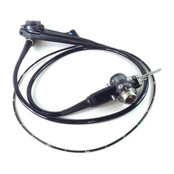
Page 16: Chapter 2 Instrument Nomenclature And Specifications
Chapter 2 Instrument Nomenclature and Specifications Chapter 2 Instrument Nomenclature and Specifications Nomenclature GIF-XP160, GIF-160, GIF-Q160 Universal cord 1. Suction connector 5. Electrical connector 2. S-cord connector mount Air pipe 3. Air supply connector 3. Water supply connector Light guide Product name and serial number Electrical contacts 4. - Page 17 Chapter 2 Instrument Nomenclature and Specifications 9. Air/water valve (MH-438) 8. Suction valve (MH-443) 7. UP/DOWN angulation lock 6. UP/DOWN angulation control knob 16. RIGHT/LEFT angulation control knob Control section 15. RIGHT/LEFT angulation lock Suction cylinder Air/water cylinder Grip section 14.
- Page 18 Chapter 2 Instrument Nomenclature and Specifications GIF-1TQ160, CF-Q160L/I, CF-Q160S Universal cord 17. Auxiliary water inlet 1. Suction connector 5. Electrical connector Auxiliary water inlet cap (MAJ-215) Air pipe 2. S-cord connector mount 3. Air supply connector 3. Water supply connector Light guide Product name and serial number Electrical contacts...
- Page 19 Chapter 2 Instrument Nomenclature and Specifications 9. Air/water valve (MH-438) 8. Suction valve (MH-443) 7. UP/DOWN angulation lock 6. UP/DOWN angulation control knob 16. RIGHT/LEFT angulation control knob Control section 15. RIGHT/LEFT angulation lock Suction cylinder Air/water cylinder Grip section 14.
- Page 20 Chapter 2 Instrument Nomenclature and Specifications GIF-XTQ160 Universal cord 17. Auxiliary water inlet 1. Suction connector 5. Electrical connector Auxiliary water inlet cap (MAJ-215) Air pipe 2. S-cord connector mount 3. Air supply connector 3. Water supply connector Light guide Product name and serial number Electrical contacts 4.
- Page 21 Chapter 2 Instrument Nomenclature and Specifications 9. Air/water valve (MH-438) 8. Suction valve (MH-443) 7. UP/DOWN angulation lock 6. UP/DOWN angulation control knob 16. RIGHT/LEFT angulation control knob Suction cylinder Control section 15. RIGHT/LEFT angulation lock Air/water cylinder Grip section Biopsy valve 14.
- Page 22 Chapter 2 Instrument Nomenclature and Specifications CF-Q160AL/I Universal cord 17. Auxiliary water inlet 1. Suction connector 5. Electrical connector Auxiliary water inlet cap (MAJ-215) Air pipe 2. S-cord connector mount 3. Air supply connector 3. Water supply connector Light guide Product name and serial number Electrical contacts 4.
- Page 23 Chapter 2 Instrument Nomenclature and Specifications 9. Air/water valve (MH-438) 8. Suction valve (MH-443) 7. UP/DOWN angulation lock 6. UP/DOWN angulation control knob 16. RIGHT/LEFT angulation control knob Control section 15. RIGHT/LEFT angulation lock Suction cylinder Air/water cylinder Grip section 14.
- Page 24 Chapter 2 Instrument Nomenclature and Specifications PCF-160AL/I Universal cord 1. Suction connector 5. Electrical connector 2. S-cord connector mount Air pipe 3. Air supply connector 3. Water supply connector Light guide Product name and serial number Electrical contacts 4. Endoscope connector EVIS EXERA GIF/CF/PCF TYPE 160 Series OPERATION MANUAL...
- Page 25 Chapter 2 Instrument Nomenclature and Specifications 9. Air/water valve (MH-438) 8. Suction valve (MH-443) 7. UP/DOWN angulation lock 6. UP/DOWN angulation control knob 16. RIGHT/LEFT angulation control knob Control section 15. RIGHT/LEFT angulation lock Suction cylinder Air/water cylinder Grip section 14.
-
Page 26: Endoscope Functions
This connector connects the endoscope to the suction tube of the suction pump. 2. S-cord connector mount This mount connects the endoscope with the Olympus electrosurgical unit via the S-cord. The S-cord conducts leakage current from the endoscope to the electrosurgical unit. To connect the S-cord, refer to the instruction manual for the electrosurgical unit. - Page 27 Chapter 2 Instrument Nomenclature and Specifications 10. Instrument channel port The instrument channel port functions as: − channel for the insertion of endo-therapy accessories − suction channel − fluid feed channel (from a syringe via the biopsy valve) 11. Insertion tube limit mark This mark shows the maximum point to which the endoscope may be inserted into the patient’s body.
-
Page 28: Specifications
Chapter 2 Instrument Nomenclature and Specifications 18. Flexibility adjustment ring (for CF-Q160AL/I, PCF-160AL/I only) Turn this ring to adjust the flexibility of the insertion tube. When the “ ” mark on the ring is aligned with the “ ” mark at the bottom of the grip section, the insertion tube has the softest condition. - Page 29 Chapter 2 Instrument Nomenclature and Specifications Specifications Endoscope functions Model GIF-XP160 Optical Field of view 120° system Direction of view Forward viewing Depth of field 3 – 100 mm Insertion tube Distal end outer ø 5.9 mm diameter Distal end enlarged 1.
- Page 30 Chapter 2 Instrument Nomenclature and Specifications Model GIF-160 GIF-Q160 Optical Field of view 140° system Direction of view Forward viewing Depth of field 3 – 100 mm Insertion tube Distal end outer ø 8.6 mm ø 9.8 mm diameter Distal end enlarged 1.
- Page 31 Chapter 2 Instrument Nomenclature and Specifications Model GIF-1TQ160 Optical Field of view 140° system Direction of view Forward viewing Depth of field 3 – 100 mm Insertion Distal end outer ø 10.9 mm tube diameter Distal end enlarged 1. Air/water nozzle 2.
- Page 32 Chapter 2 Instrument Nomenclature and Specifications Model GIF-XTQ160 Optical Field of view 140° system Direction of view Forward viewing Depth of field 3 – 100 mm Insertion Distal end outer ø 12.9 mm tube diameter Distal end enlarged 1. Air/water nozzle 2.
- Page 33 Chapter 2 Instrument Nomenclature and Specifications Model CF-Q160L/I CF-Q160S Optical Field of view 140° system Direction of view Forward viewing Depth of field 3 – 100 mm Insertion tube Distal end outer ø 12.8 mm diameter Distal end 1. Air/water nozzle enlarged 2.
- Page 34 Chapter 2 Instrument Nomenclature and Specifications Model CF-Q160AL/I PCF-160AL/I Optical Field of view 140° system Direction of view Forward viewing Depth of field 3 – 100 mm 3 – 100 mm Insertion tube Distal end outer ø 12.8 mm ø 11.3 mm diameter Distal end enlarged 1.
- Page 35 Chapter 2 Instrument Nomenclature and Specifications Medical This device complies with the Device requirements of Directive 93/42/EEC Directive concerning medical devices. Classification: Class II a Applied standard; This instrument complies with the IEC 60601-1-2: 2001 standards listed in the left column. CISPR 11 of emission: Group 1, Class B This instrument complies with the EMC...
-
Page 36: Chapter 3 Preparation And Inspection
Should the slightest irregularity be suspected, do not use this instrument and see Chapter 5, “Troubleshooting”. If the irregularity is still suspected after consulting Chapter 5, contact Olympus. Damage or irregularity may compromise patient or user safety and may result in more severe equipment damage. -
Page 37: Preparation Of The Equipment
Chapter 3 Preparation and Inspection Preparation of the equipment Prepare the equipment shown in Figure 3.1 (for compatibility, see the “System chart” in the Appendix) and personal protective equipment, such as eye wear, face mask, moisture-resistant clothing and chemical-resistant gloves, before each use. -
Page 38: Inspection Of The Endoscope
Chapter 3 Preparation and Inspection Inspection of the endoscope Clean and disinfect or sterilize the endoscope as described in its companion reprocessing manual. Then remove the water-resistant cap from the endoscope connector. Inspection of the endoscope Inspect the control section and the endoscope connector for excessive scratching, deformation, loose parts or other irregularities. - Page 39 Chapter 3 Preparation and Inspection Using both hands, bend the insertion tube of the endoscope into a semicircle. Then, moving your hands as shown by the arrows in Figure 3.3, confirm that the entire insertion tube can be smoothly bent to form a semicircle and that the insertion tube is pliable.
- Page 40 Chapter 3 Preparation and Inspection Inspection of the flexibility adjustment mechanism (for CF-Q160AL/I, PCF-160AL/I only) Confirm that the index markings (“ ”, “1”, “2”, “3”) on the flexibility adjustment ring and the “ ” mark at the bottom of the grip section are clearly visible (see Figure 3.4).
- Page 41 Chapter 3 Preparation and Inspection Figure 3.5 Inspection of the bending mechanisms Perform the following inspections while the bending section is straight. If the movement of the UP/DOWN angulation lock, RIGHT/LEFT angulation lock and their angulation control knobs are loose and/or not smooth, or the bending section does not angulate smoothly, the bending mechanism may be abnormal.
- Page 42 Chapter 3 Preparation and Inspection Figure 3.6 Inspection of the UP/DOWN angulation mechanism Move the UP/DOWN angulation lock all the way in the opposite direction of the “F ” mark. Then turn the UP/DOWN angulation control knob in the “ U”...
-
Page 43: Preparation And Inspection Of Accessories
Chapter 3 Preparation and Inspection Preparation and inspection of accessories Clean and disinfect or sterilize the air/water valve, suction valve, biopsy valve and auxiliary water tube (for endoscopes with auxiliary water feeding only) as described in the endoscope’s companion reprocessing manual. Inspection of the air/water and suction valves Confirm that the holes of the valves are not blocked (see Figures 3.7 and 3.8). - Page 44 Chapter 3 Preparation and Inspection • The air/water and suction valves are consumables. If the inspection of the air/water or suction valve reveals any irregularities, use new valves. • Only air/water valve MH-438 and suction valve MH-443 should be used with this endoscope. Inspection of the biopsy valve The biopsy valve is a consumable item that should be inspected before each use.
- Page 45 Chapter 3 Preparation and Inspection Slit Main body Figure 3.10 Inspection of the auxiliary water inlet cap (for endoscopes with auxiliary water feeding only) Inspect the auxiliary water inlet cap for dents, cracks or other irregularities. Inspection of the auxiliary water tube (for endoscopes with auxiliary water feeding only) Inspect the auxiliary water tube for cracks, scratches, flaws and other damage (see Figure 3.11).
- Page 46 Chapter 3 Preparation and Inspection Inspection of the mouthpiece (for GIF models only) Do not use a mouthpiece that is damaged, deformed or reveals other irregularities. Doing so may cause patient injury and/or equipment damage. Placing the mouthpiece in the patient’s mouth before the procedure prevents the patient from biting and/or damaging the endoscope’s insertion tube.
-
Page 47: Attaching Accessories To The Endoscope
Chapter 3 Preparation and Inspection Attaching accessories to the endoscope The air/water valve and the suction valve do not require lubrication. Lubricants can cause swelling of the valves’ seals, which will impair valve function. Attaching the suction valve Align the two metal ridges on the underside of the suction valve with the two holes in the suction cylinder. - Page 48 Chapter 3 Preparation and Inspection Attaching the air/water valve Attach the air/water valve to the air/water cylinder of the endoscope (see Figure 3.14). Confirm that the valve fits properly without any bulging of the skirt. Air/water valve Suction valve Skirt Suction cylinder Air/water cylinder Figure 3.14...
-
Page 49: Inspection And Connection Of Ancillary Equipment
Chapter 3 Preparation and Inspection Inspection and connection of ancillary equipment Inspection of ancillary equipment • Attach the water container to the specified receptacle on the trolley or the light source. If the water container is attached anywhere else, water may drip from the water container’s water supply tube, and equipment malfunction can result. - Page 50 Chapter 3 Preparation and Inspection Connect the water container’s connection adapter to the air supply connector and water supply connector (see Figure 3.16). Air supply connector Water supply connector Water container’s connection adapter Endoscope connector Figure 3.16 Align the mark on the videoscope cable EXERA or the videoscope cable 100 with mark 1 on the endoscope connector and push it in until it stops (see Figure 3.17).
- Page 51 Chapter 3 Preparation and Inspection Connect the suction tube from the suction pump to the suction connector on the endoscope connector (see Figure 3.18). Suction pump Suction tube Figure 3.18 Open the auxiliary water inlet cap (for endoscopes with auxiliary water feeding only, see Figure 3.19). Connect the auxiliary water tube to the auxiliary water inlet on the endoscope connector and turn it clockwise until it stops (for endoscopes with auxiliary water feeding only, see Figure 3.19).
-
Page 52: Inspection Of The Endoscopic System
Chapter 3 Preparation and Inspection Inspection of the endoscopic system Inspection of the endoscopic image Turn ON the video system center, light source and video monitor and inspect the endoscopic image as described in their respective instruction manuals. If the object cannot be seen clearly, wipe the objective lens using a clean, lint-free cloth moistened with 70% ethyl or isopropyl alcohol. - Page 53 If this fails to stop air bubbles from being emitted, do not use the endoscope, as there may be a malfunction. Contact Olympus. When the distal end of the insertion tube is immersed less than 10 cm below the surface of the sterile water, a small amount of air bubbles may be emitted from the air/water nozzle even when the air/water valve is not operated.
- Page 54 If the reattached or replaced suction valve fails to operate smoothly, the endoscope may be malfunctioning; stop using it and contact Olympus. • If the biopsy valve leaks, replace it with a new one. A leaking biopsy valve can reduce the efficacy of the endoscope’s...
- Page 55 Chapter 3 Preparation and Inspection Inspection of the instrument channel Keep your eyes away from the distal end when inserting endo-therapy accessories. Extending the endo-therapy accessory from the distal end could cause eye injury. Insert the endo-therapy accessory through the biopsy valve. Confirm that the endo-therapy accessory extends smoothly from the distal end.
- Page 56 Chapter 3 Preparation and Inspection If the auxiliary water channel is used for feeding water, never disconnect the auxiliary water tube during an examination; leave it attached until the endoscope is precleaned. If the auxiliary water tube is detached before precleaning, water remaining in the auxiliary water channel may be spilled on the surrounding equipment.
-
Page 57: Chapter 4 Operation
Chapter 4 Operation Chapter 4 Operation The operator of this instrument must be a physician or medical personnel under the supervision of a physician and must have received sufficient training in clinical endoscopic technique. This manual, therefore, does not explain or discuss clinical endoscopic procedures. - Page 58 Chapter 4 Operation • Never insert or withdraw the endoscope under any of the following conditions. Otherwise, patient injury can result. − Insertion or withdrawal while the endo-therapy accessory extends from the distal end of the endoscope. − Insertion or withdrawal while the bending section is locked in position.
- Page 59 Chapter 4 Operation • The flexibility of the insertion tube of the CF-Q160AL/I can be adjusted to less than, equal to or more than that of the CF-Q140L/I. The flexibility of the insertion tube of the PCF-160AL/I can be adjusted in equal to or more than that of the PCF-140L/I.
-
Page 60: Insertion
Chapter 4 Operation Insertion Holding and manipulating the endoscope The control section of the endoscope is designed to be held in the left hand. The air/water and suction valves can be operated using the left index finger. The UP/DOWN angulation control knob can be operated using the left thumb. The right hand is free to manipulate the insertion tube and the RIGHT/LEFT angulation control knob (see Figure 4.1). - Page 61 Chapter 4 Operation > 10 cm Figure 4.2 If necessary, apply a medical-grade, water-soluble lubricant to the insertion tube. Place the mouthpiece between the patient’s teeth or gums, with the outer flange on the outside of the patient’s mouth (for GIF models only). Insert the insertion tube of the endoscope into the splinting tube if required, and apply the lubricant to it (for CF/PCF models only).
- Page 62 Chapter 4 Operation Angulation of the distal end Operate the angulation control knobs as necessary to guide the distal end for insertion and observation. The endoscope’s angulation locks are used to hold the angulated distal end in position. • When passing an endo-therapy accessory through the instrument channel while the angulation is locked, the angle of the distal end may change.
- Page 63 Chapter 4 Operation Before inserting or withdrawing the endoscope, set the insertion tube to an appropriate level of flexibility by turning the flexibility adjustment ring as required (see Figure 4.3). Stiffer Softer Figure 4.3 When changing the insertion tube’s flexibility during a procedure, turn the flexibility adjustment ring slowly, and closely monitor the position of flexibility index marking, the endoscopic image and the patient’s condition.
- Page 64 Chapter 4 Operation Air/water feeding If the sterile water level in the water container is too low, then air, not water, will be supplied. In this case, turn OFF the airflow regulator on the light source and add more sterile water to the water container.
- Page 65 Chapter 4 Operation • When aspirating, maintain the suction pressure at the lowest level necessary to perform the procedure. Excessive suction pressure could cause aspiration of and/or injury to the mucous membrane. In addition, patient fluids could leak or spray from the biopsy valve, posing an infection-control risk. •...
-
Page 66: Using Endo-Therapy Accessories
Chapter 4 Operation When disconnecting the syringe or the water tube from the water pump during examination, disconnect it directly from the luer port but leave the auxiliary water tube itself attached. Observation of the endoscopic image Refer to the light source’s instruction manual for instructions on how to adjust the brightness. - Page 67 Chapter 4 Operation • If the distal end of an endo-therapy accessory is not visible in the endoscopic image, do not open the distal end or extend the needle of the instrument. This could cause patient injury, bleeding, perforation and/or equipment damage. •...
- Page 68 Chapter 4 Operation • Hold the part which is close to the biopsy valve of the endo-therapy accessory, and insert it straight, slowly, and little by little to the biopsy valve. Otherwise, the endo-therapy accessory and/or biopsy valve could be damaged. This can reduce the efficacy of the endoscope’s suction system, and may leak or spray patient debris or fluids, posing an infection-control risk.
- Page 69 Chapter 4 Operation Withdrawal of endo-therapy accessories • Do not withdraw the endo-therapy accessory if the tip is open or extended from its sheath; patient injury and/or instrument damage may occur. • Withdraw the endo-therapy accessory slowly and straight out of the biopsy valve.
- Page 70 Chapter 4 Operation • Not all parts of the endoscope are electrically insulated. When applying high frequency current, there is a danger of unintentional diathermy burns. Always wear electrically insulating chemical-resistant gloves. • To avoid patient injury and/or damage to the endoscope, never emit high frequency current before confirming that the electrode section of the high frequency endo-therapy accessory is extended from the distal end of the endoscope...
-
Page 71: Withdrawal Of The Endoscope
If the endoscope cannot be withdrawn from the patient smoothly, do not attempt to forcibly withdraw it; leave it inside the patient and immediately contact Olympus. Forcibly withdrawing the endoscope may cause patient injury. Aspirate accumulated air, blood, mucus or other debris by depressing the suction valve. -
Page 72: Transportation Of The Endoscope
Chapter 4 Operation Transportation of the endoscope Transporting within the hospital Set the insertion tube to the softest condition (for CF-Q160AL/I, PCF-160AL/I only). When carrying the endoscope with the auxiliary water tube connected to the auxiliary water inlet, attach the clip of the auxiliary water tube to the universal cord (for endoscopes with the auxiliary water feeding only, see Figure 4.5). - Page 73 Chapter 4 Operation Transporting outside the hospital Transport the endoscope in the carrying case. Always clean, disinfect or sterilize the endoscope after removing it from the carrying case. • The carrying case cannot be cleaned, disinfected or sterilized. Clean and disinfect or sterilize the endoscope before placing it in the carrying case.
-
Page 74: Chapter 5 Troubleshooting
Some problems that appear to be malfunctions may be correctable by referring to Section 5.1, “Troubleshooting guide”. If the problem cannot be resolved by the described remedial action, stop using the endoscope and send it to Olympus for repair. Olympus does not repair accessory parts. If an accessory part becomes damaged, contact Olympus to purchase a replacement. - Page 75 Chapter 5 Troubleshooting Endoscope functions Angulation Irregularity Possible cause Solution description Resistance is The angulation lock(s) is Rotate angulation lock(s) in the encountered when (are) engaged. “F ” direction. rotating angulation control knob(s). Air/water feeding Irregularity Possible cause Solution description No air feeding.
- Page 76 Chapter 5 Troubleshooting Suction Irregularity Possible cause Solution description The suction is absent or The biopsy valve is not Attach it correctly. insufficient. attached properly. The biopsy valve is Replace it with a new one. damaged. The suction pump is not Adjust the suction pump’s setting set properly.
- Page 77 Chapter 5 Troubleshooting Flexibility adjustment (for CF-Q160AL/I, PCF-160AL/I only) Irregularity Possible cause Solution description Too difficult to turn the The insertion tube is Straighten the insertion tube. flexibility adjustment looped. ring. Auxiliary water feeding (for endoscopes with auxiliary water feeding only) Irregularity Possible cause Solution...
-
Page 78: Withdrawal Of The Endoscope With An Abnormality
If the endoscope or endo-therapy accessory cannot be withdrawn from the patient smoothly, do not attempt to forcibly withdraw it. If any irregularities are suspected, immediately contact Olympus. Forcibly withdrawing the endoscope or endo-therapy accessory may cause patient injury, bleeding and/or perforation. - Page 79 Chapter 5 Troubleshooting When the endoscopic image does not appear on the monitor or the frozen image cannot be restored Turn OFF all equipment except the video system center, the light source and the monitor. Turn the video system center and light source OFF and then ON again. If the endoscopic image appears or the frozen image is restored, follow the procedure of Step 2.
-
Page 80: Returning The Endoscope For Repair
Olympus. Olympus is not liable for any injury or damage which occurs as a result of repairs attempted by non-Olympus personnel. Before returning the endoscope for repair, contact Olympus. With the... -
Page 81: Appendix
New products released after the introduction of this instrument may also be compatible for use in combination with this instrument. For further details, contact Olympus. If combinations of equipment other than those shown below are used, the full responsibility is assumed by the medical treatment facility. - Page 82 Appendix GIF-XP160 Suction pumps GIF-160 GIF-Q160 GIF-1TQ160 GIF-XTQ160 CF-Q160L/I Auxiliary water tube CF-Q160AL/I (MAJ-855 for endoscopes with PCF-160AL/I auxiliary water feeding only) CF-Q160S KV-4 KV-5 SSU-2 ∗ Videoscope cable Videoscope cable 100 ∗ ∗ EXERA (MAJ-843) (MH-976 , MD-148 Sclerotherapy balloon (for GIF models only) ∗...
- Page 83 Appendix Biopsy valve (MB-358) Forceps suction plug (T-Plug) (MH-405 for Endo-therapy accessories GIF-XTQ160 only) Electrosurgical units See next page. Electrosurgical accessories UES-20/30 See next page. PSD-20/30 Sclerotherapy tube (for GIF models only) Cleaning and disinfection equipment Auxiliary water tube (MAJ-855 for endoscopes Ultrasonic cleaner with auxiliary water (KS-2/ENDOSONIC)
- Page 84 Appendix EVIS EXERA video system center/EVIS video system centers Videoscope cable Videoscope cable 100 EXERA Endoscope CV-100 CV-140 CV-160 – – GIF-XP160 GIF-160 – GIF-Q160 – GIF-1TQ160 – GIF-XTQ160 – CF-Q160L/I – CF-Q160AL/I PCF-160AL/I – CF-Q160S applicable – not applicable Accessories (for GIF models only) Sclerotherapy Forceps...
- Page 85 Appendix Endo-therapy accessories BIOPSY FORCEPS BIOPSY FORCEPS (Fenestrated) Single side open With needle Alligator jaws Standard Endoscope GIF-XP160 – – FB-15K-1 FB-19K-1/21K-1 GIF-160 FB-11K-1 – – FB-25K-1 GIF-Q160 FB-11K-1 – – FB-25K-1 GIF-1TQ160 FB-11K-1 FB-13K-1 FB-15K-1 FB-25K-1 GIF-XTQ160 FB-11K-1 FB-13K-1 FB-15K-1 FB-25K-1 CF-Q160L...
- Page 86 Appendix ROTATABLE BIOPSY FORCEPS (Fenestrated) BIOPSY FORCEPS (Fenestrated) Alligator jaws and Alligator jaws and Alligator jaws and rat tooth with rat tooth rat tooth needle Standard type (Swinging type/ (Swinging type) (Swinging type/ Elongated cups) Elongated cups) Endoscope GIF-XP160 – FB-52K-1 –...
- Page 87 Appendix ROTATABLE BIOPSY FORCEPS (Fenestrated) Alligator jaws and Alligator jaws and Alligator jaws and rat tooth with Elongated cups rat tooth rat tooth needle with needle (Swinging type/ (Swinging type) (Swinging type/ Elongated cups) Elongated cups) Endoscope GIF-XP160 – – –...
- Page 88 Appendix DISPOSABLE BIOPSY FORCEPS Alligator jaws-step Alligator jaws-step Oval Oval with needle with needle Endoscope GIF-XP160 FB-211K FB-221K FB-231K FB-241K GIF-160 FB-210K FB-220K FB-230K FB-240K GIF-Q160 FB-210K FB-220K FB-230K FB-240K GIF-1TQ160 FB-212U FB-222U FB-232U FB-242U GIF-XTQ160 FB-212U FB-222U FB-232U FB-242U CF-Q160L FB-212U FB-222U...
- Page 89 Appendix GRASPING FORCEPS Alligator jaws Rat tooth Covered tips Sharp tooth Endoscope GIF-XP160 – FG-14P-1 FG-20P-1 – FG-8L-1/48L-1/ GIF-160 FG-6L-1 FG-21L-1 FG-32L-1 50L-1 FG-8L-1/48L-1/ GIF-Q160 FG-6L-1 FG-21L-1 FG-32L-1 50L-1 FG-9L-1/48L-1/ GIF-1TQ160 FG-7L-1 FG-21L-1 FG-32L-1 50L-1 FG-9L-1/48L-1/ GIF-XTQ160 FG-7L-1 FG-21L-1 FG-32L-1 50L-1 CF-Q160L FG-7U-1...
- Page 90 Appendix GRASPING FORCEPS Rat tooth with W shape jaw Basket type Tripod type alligator jaws Endoscope GIF-XP160 – FG-4L-1 FG-17K-1 – FG-42L-1/47L-1/ GIF-160 FG-4L-1 FG-16L-1 FG-45L-1 49L-1 FG-42L-1/47L-1/ GIF-Q160 FG-4L-1 FG-16L-1 FG-45L-1 49L-1 FG-42L-1/47L-1/ GIF-1TQ160 FG-4L-1 FG-16L-1 FG-45L-1 49L-1 FG-42L-1/47L-1/ GIF-XTQ160 FG-4L-1 FG-16L-1...
- Page 91 Appendix SINGLE USE GRASPING GRASPING SURGICAL FORCEPS LOOP CUTTER FORCEPS SCISSORS Pentapod type Tripod type Endoscope GIF-XP160 – – – – GIF-160 FG-46L-1 FG-600U FS-3L-1 FS-5L-1 GIF-Q160 FG-46L-1 FG-600U FS-3L-1 FS-5L-1 GIF-1TQ160 FG-46L-1 FG-600U FS-3L-1 FS-5L-1 GIF-XTQ160 FG-46L-1 FG-600U FS-3L-1 FS-5L-1 CF-Q160L FG-46U-1...
- Page 92 Appendix CLIP FIXING DISPOSABLE CLIP LIGATING DEVICE DEVICE FIXING DEVICE Endoscope GIF-XP160 – – – – ∗ GIF-160 HX-5LR-1 HX-200L-135 HX-20L-1 HX-21L-1 ∗ GIF-Q160 HX-5LR-1 HX-200L-135 HX-20L-1 HX-21L-1 ∗ GIF-1TQ160 HX-6UR-1 HX-200L-135 HX-20L-1 HX-21L-1 ∗ GIF-XTQ160 HX-6UR-1 HX-200L-135 HX-20L-1 HX-21L-1 CF-Q160L HX-6UR-1 HX-200U-135...
- Page 93 Appendix DISTAL ATTACHMENT Straight Oblique Straight with rim Oblique with rim Endoscope GIF-XP160 – – – – ∗ ∗ ∗ ∗ GIF-160 MH-462 MH-587 MH-593 MAJ-289 ∗ ∗ ∗ ∗ GIF-Q160 MH-463 MH-588 MH-594 MAJ-290 ∗ ∗ ∗ ∗ GIF-1TQ160 MH-464 MH-589 MH-595...
- Page 94 Appendix Electrosurgical accessories POLYPECTOMY SNARE Crescent Hexagonal Oval Mini-oval Endoscope GIF-XP160 SD-7P-1 SD-8P-1 – – GIF-160 SD-5L-1 SD-6L-1 SD-9L-1/11L-1 SD-12L-1/13L-1 GIF-Q160 SD-5L-1 SD-6L-1 SD-9L-1/11L-1 SD-12L-1/13L-1 GIF-1TQ160 SD-5L-1 SD-6L-1 SD-9L-1/11L-1 SD-12L-1/13L-1 GIF-XTQ160 SD-5L-1 SD-6L-1 SD-9L-1/11L-1 SD-12L-1/13L-1 CF-Q160L SD-5U-1 SD-6U-1 SD-9U-1/11U-1 SD-12U-1/13U-1 CF-Q160I SD-5U-1 SD-6U-1...
- Page 95 Appendix DISPOSABLE POLYPECTOMY Extra mini-oval Crescent Oval (With spiral) Endoscope GIF-XP160 – SD-221L-25 – GIF-160 SD-210L-10 SD-221L-25 SD-230U-20 GIF-Q160 SD-210L-10 SD-221L-25 SD-230U-20 GIF-1TQ160 SD-210L-10 SD-221L-25 SD-230U-20 GIF-XTQ160 SD-210L-10 SD-221L-25 SD-230U-20 CF-Q160L SD-210U-10 SD-221U-25 SD-230U-20 CF-Q160I SD-210U-10 SD-221U-25 SD-230U-20 CF-Q160AL SD-210U-10 SD-221U-25 SD-230U-20 CF-Q160AI...
-
Page 96: Emc Information
Appendix EMC information This model is intended for use in the electromagnetic environments specified below. The user and the medical staff should ensure that it is used only in these environments. Magnetic emission compliance information and recommended electromagnetic environments Emission standard Compliance Guidance RF emissions... - Page 97 Appendix Electromagnetic immunity compliance information and recommended electromagnetic environments IEC 60601-1-2 Immunity test Compliance level Guidance test level Electrostatic Contact: Same as left Floors should by be made of wood, concrete, ±2, ±4, ±6 kV discharge (ESD) or ceramic tile that hardly produces static. If floors are covered with synthetic material that IEC 61000-4-2 Air:...
- Page 98 Appendix Cautions and recommended electromagnetic environment regarding portable and mobile RF communications equipment such as cellular phones IEC 60601-1-2 Compliance Immunity test Guidance test level level Formula for recommended separation distance =3 according to the compliance level) Conducted RF 3 Vrms 3 V (V ------ - (150 kHz –...
- Page 99 Appendix Recommended separation distance between portable and mobile RF communications equipment and this instrument Separation distance according to frequency of transmitter (m) (calculated as V =3 and E Rated maximum output 150 kHz – 80 MHz 80 MHz – 800 MHz 800 MHz –...
- Page 101 ©2003 OLYMPUS MEDICAL SYSTEMS CORP. All rights reserved. No part of this publication may be reproduced or distributed without the express written permission of OLYMPUS MEDICAL SYSTEMS CORP. OLYMPUS is a registered trademark of OLYMPUS CORPORATION.
- Page 102 Manufactured by 2951 Ishikawa-cho, Hachioji-shi, Tokyo 192-8507, Japan Fax: (042)646-2429 Telephone: (042)642-2111 Distributed by 3500 Corporate Parkway, P.O. Box 610 Center Valley, PA 18034-0610, U.S.A. Fax: (484)896-7128 Telephone: (484)896-5000 One Corporate Drive, Orangeburg, N.Y. 10962, U.S.A. Fax: (845)398-9444 Telephone: (845)398-9400 5301 Blue Lagoon Drive, Suite 290 Miami, FL 33126-2097, U.S.A.
Need help?
Do you have a question about the EVIS EXERA GIF XP160 and is the answer not in the manual?
Questions and answers