
Related Manuals for Sonoscape S2


Summary of Contents for Sonoscape S2
- Page 1 S2/S2BW Digital Color Doppler Ultrasound System S2/S2BW Digital Color Doppler Ultrasound System Basic Operator’s Manual SonoScape Co., Ltd. P/N: 4710.00149A01...
- Page 2 S2/S2BW Digital Color Doppler Ultrasound System Regulatory Requirement This product complies with regulatory requirements of the following European directive 93/42/EEC as amended by 2007/47/EC concerning medical devices. NOTE: Some options are not available on some models! This manual is subject to change without prior notice and without legal obligation attached!
- Page 3 S2/S2BW Digital Color Doppler Ultrasound System Revision History Revision Date Description June 2012 First edition. P/N: 4710.00149A01 Compiled on June, 2012. P/N: 4710.00149A01...
-
Page 4: Table Of Contents
S2/S2BW Digital Color Doppler Ultrasound System Contents Introduction System Safety and Maintenance Safety Overview ........2-1 Symbol Used . - Page 5 S2/S2BW Digital Color Doppler Ultrasound System Main User Interface ........4-25...
- Page 6 S2/S2BW Digital Color Doppler Ultrasound System CFM Mode Enter CFM Mode ........7-1 7.1.1...
- Page 7 S2/S2BW Digital Color Doppler Ultrasound System 10.1.2 Active Mode ........10-1 10.2...
- Page 8 S2/S2BW Digital Color Doppler Ultrasound System A Description of Symbols B Acoutic Output Data C Information of EU Representative P/N: 4710.00149A01...
- Page 9 S2/S2BW Digital Color Doppler Ultrasound System P/N: 4710.00149A01...
-
Page 10: Introduction
S2/S2BW is a full digital color Doppler ultrasound system for general application. The high performances of the SonoScape S2 series stem from the advanced ultrasound doppler imaging technologies that include full digital beam-former, wide dynamic range, multi-beam processing, etc. The ergonomic user-friendly design enables user to customize the system according to the specific application,... - Page 11 S2/S2BW Digital Color Doppler Ultrasound System P/N: 4710.00149A01...
-
Page 12: System Safety And Maintenance
S2/S2BW Digital Color Doppler Ultrasound System Chapter 2 System Safety and Maintenance 2.1 Safety Overview This section discusses measures to ensure the safety of both the operator and patient. To ensure the safety of both operator and patient, please read the relevant details in this chapter carefully before operating this system. -
Page 13: Symbol Used
S2/S2BW Digital Color Doppler Ultrasound System • Do not use the switch at the back of the unit for normal shut down. Always use the power-on button in the keyboard area • Do not unplug probe when the system is in active operation. Doing so may damage the probe. Always go to EXAM screen when need to remove the probe. -
Page 14: Adverse Effects And Precautions
S2/S2BW Digital Color Doppler Ultrasound System 2.4 Adverse Effects and Precautions This ultrasound system, same as all other diagnostic ultrasound system in the market, should be used only for clinically appropriate reasons, for the shorteds period of time and at power settings as low as reasonably achievable (ALARA). - Page 15 S2/S2BW Digital Color Doppler Ultrasound System 7. Do not use endo-cavity probes if there is noticeable self heating of the probe when operating in the air. Although applicable to any probe, take particular care during trans-vaginal exams during the first eight weeks of gestation.
-
Page 16: Understanding The Mi/Ti Display
S2/S2BW Digital Color Doppler Ultrasound System 2.5.1.1 Temperature Display for Transducers Intended for Internal Use For transducers intended for internal applications, e.g. the intracavitary transducers, the transducer tip temperature is displayed on the screen.To protect the patient against the harm of excessive temperature, the system automatically turns off the transducer when the temperature of the transducer reaches a threshold temperature. - Page 17 S2/S2BW Digital Color Doppler Ultrasound System Distance Frequency (MHz) (cm) 0,9332 0,8128 0,7080 0,5957 0,8710 0,6607 0,5012 0,3548 0,8128 0,5370 0,3548 0,2113 0,7586 0,4365 0,2512 0,1259 0,7080 0,3548 0,1778 0,0750 0,6607 0,2884 0,1259 0,0447 0,6166 0,2344 0,0891 0,0266 0,5754 0,1903...
-
Page 18: Environmental Requirements
S2/S2BW Digital Color Doppler Ultrasound System The threshold under optimum conditions of pulsed ultrasound is predicted by the ratio of the peak pressure to the square root of the frequency. Pr’ is the de-rated (0.3) peak rare-fractional pressure in Mpa at the point where PII is the maximum, and fc is the center frequency in MHz. -
Page 19: Electrical Requirements
Caution for using AC adapter: • Use the AC adapter approved by SonoScape only. • AC adapter can get very hot if being used in poorly ventilated area. Maintain good ventila- tion for heat dissipation. Do not put any items on the AC adapter or the power cable. -
Page 20: Electrical Safety
Other accessories or devices not listed in the supported accessories’ list (Contact the manufacturer for the updated and complete list of accessories.) must not be used. 3. Installation of the system must be performed by the service engineer authorized by SonoScape. P/N: 4710.00149A01... -
Page 21: Transducer Maintenance
S2/S2BW Digital Color Doppler Ultrasound System 2.9 Transducer Maintenance The transducers/probes provided with the system are durable and have reliable performance. These precision instruments should be inspected daily and handled with care. Please observe the following PRECAUTIONS: • Do not drop the transducer on the hard surface. This can damage the transducer elements and compromise the electrical safety of the transducer. - Page 22 3) You should consult the manufacturer’s instructions to prepare, store and use the disinfectant. Consult SonoScape if you are not sure about the compatibility of these agents with the probes. Many of the chemical disinfectants are potentially toxic and many require adequate precautions, such as proper ventilation, personal protective devices (gloves, face/eye protection, etc.) and rinsing before reusing the...
- Page 23 S2/S2BW Digital Color Doppler Ultrasound System 3) Users should be aware of latex-sensitivity issues and have available nonlatex-containing barriers. 4. ASEPTIC TECHNIQUE 1. For the protection of the patient and the health care worker, all endocavitary examinations should be performed by the operator wearing gloves properly throughout the ultrasound diagnostic process.
-
Page 24: System Transportation
S2/S2BW Digital Color Doppler Ultrasound System 2.10 System Transportation 2.10.1 Moving the System When moving or transporting the system, take the PRECAUTIONS listed below. Before moving the system: 1. Completely power off the system. 2. Unplug the power cord. 3. Disconnect all cables attached to the system. - Page 25 S2/S2BW Digital Color Doppler Ultrasound System P/N: 4710.00149A01 2-14...
-
Page 26: System Specifications
S2/S2BW is a full digital color Doppler ultrasound system for general application. Thanks to the unique full digital construction, VLSI and computer platform based on Linux system, S2/S2BW can maximize the use of its transducers and enhance the diagnosis effect; besides high performance, it is also flexible and lightweight. -
Page 27: Base System
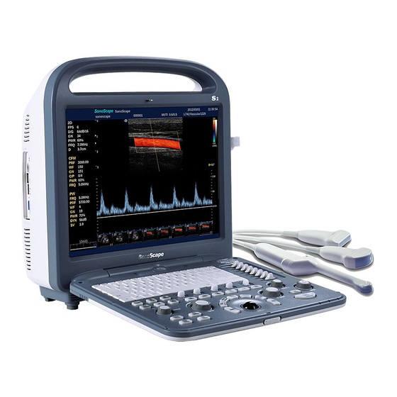
S2/S2BW Digital Color Doppler Ultrasound System 3.2 Base System 3.2.1 Front View P/N: 4710.00149A01... -
Page 28: Side View
S2/S2BW Digital Color Doppler Ultrasound System 3.2.2 Side View Figure 3.1: Side view Audio port Video port USB port DICOM port VGA port P/N: 4710.00149A01... -
Page 29: Rear View
S2/S2BW Digital Color Doppler Ultrasound System 3.2.3 Rear View Figure 3.2: Rear view Transducer socket port 1 Video port Transducer socket port 2 ECG port Printer port Equipotential point(earth) Foot switch Power interface P/N: 4710.00149A01... -
Page 30: Keyboard Layout
S2/S2BW Digital Color Doppler Ultrasound System 3.2.4 Keyboard Layout Figure 3.3: Keyboard Layout 3.2.5 Keyboard Description Description EXAM return the system to the preparation mode (EXAM screen). Patient access the Patient Information interface. Dual activate the dual display format. flip the ultrasound image left/right. - Page 31 S2/S2BW Digital Color Doppler Ultrasound System Description of the Usage Annotation insert texts or predefined annotation items. Bodymark insert Bodymarks. Arrow insert an arrow symbol on the ultrasound image 3D/4D start volume mode (3D or real time 4D mode). m-Tuning press this key during scanning to let the system automatically adjust various parameters to obtain high quality image.
- Page 32 S2/S2BW Digital Color Doppler Ultrasound System Description of the Usage activate the Tissue Harmonic Imaging. turn on/off Color Doppler Imaging. turn on/off Doppler Power Imaging. Turn on/off Power Doppler imaging. Turn on/off Continuous Doppler imaging. M Mode turn on/off M mode imaging.
-
Page 33: Probes And Accessories
S2/S2BW Digital Color Doppler Ultrasound System Description of the Usage this key serves as the confirmation key. to set, fixate markers and activate buttons/items marked by the cursor. ZOOM zoom ultrasound image. DISTANCE measure the distance between two points. TRACE press this key and move the trackball to draw a trace on the ultrasound image;... -
Page 34: Starting The System
S2/S2BW Digital Color Doppler Ultrasound System Chapter 4 Starting the System 4.1 Probe Connection Attention! • Prior to connecting or disconnecting a probe freeze the image or back to EXAM screen. • It is unnecessary to switch the unit off. - Page 35 AC power supply available, it is always recommended to use the AC supply instead of the battery. Warning! • The AC adapter is specifically designed for the SonoScape S2 ultrasound system, do not use it with other equipments. To avoid electric hazard and system damage, use only the AC adapter approved by SonoScape with the S2 ultrasound system.
-
Page 36: Using Battery
• Do not short the metal pins on the connectors using metal objects. Information for disposing the battery: To dispose and recycle the ultrasound system and the battery properly, contact SonoScape representative for instructions. Turn on the system without connecting the AC adapter to use the battery as the source. -
Page 37: Led Indicators
S2/S2BW Digital Color Doppler Ultrasound System 2. Attach the connector of the power cable to the AC adapter firmly. 3. Connect the power plug of the AC adapter to the outlet marked with “hospital grade”. 4. Connect the DC output plug into the power supply socket on the ultrasound system. The system automatically charges the battery and takes the AC supply as source. -
Page 38: Patient Information
S2/S2BW Digital Color Doppler Ultrasound System Using AC power supply. System can be on or off. Indications Battery full Green Green Battery not full and being charged. Yellow Green Table 4.1: Status of the power supply - using AC source Using battery. - Page 39 S2/S2BW Digital Color Doppler Ultrasound System Figure 4.3: Exam Review window If no examination is active, the Patient Review screen will be displayed. P/N: 4710.00149A01...
-
Page 40: Create New Patient
S2/S2BW Digital Color Doppler Ultrasound System Figure 4.4: Patient Review window Note When this ultrasound system is connected to a worklist server (e.g., HIS/RIS), you may select a patient from a list of patients. For detailed instructions, refer to “Acquire Patient Information from Worklist Server”. - Page 41 S2/S2BW Digital Color Doppler Ultrasound System Figure 4.5: New Patient window Patient ID This item is required, however, can be generated automatically by the sytem. Maximum of 64 characters allowed. Last Name Enter the last name of the patient. First Name Enter the first name of the patient.
- Page 42 S2/S2BW Digital Color Doppler Ultrasound System Note • Click to save the patient information. The patient ID will no longer be modifiable. After saving images or cine clips for this patient, the name of the patient will also become unmodifiable.
- Page 43 S2/S2BW Digital Color Doppler Ultrasound System 5. When multiple exam cases have been performed for a patient, you can select multiple exam cases and click Group Case to load all exam cases to the local system. Note Patients list can be loaded only when valid DICOM worklist server addresses have been correctly provided.
- Page 44 S2/S2BW Digital Color Doppler Ultrasound System 4.3.1.2 Exam Type – Obstetrical (OB) Obstetrical Exam Information Date Select LMP or IVF and enter the corresponding date. Click to select the estimated date of confinement. Age(wks/days) Gestational age should be less than 44 weeks and 6 days.
- Page 45 S2/S2BW Digital Color Doppler Ultrasound System Note Date of LMP/IVF, EDC and gestational age can be calculated from each other. Enter one of them and the other two would be calculated automatically. When LMP is selected, • days • Current date days •...
- Page 46 S2/S2BW Digital Color Doppler Ultrasound System Menopausal Check the box for menopausal patient. 4.3.1.4 Exam Type – Cardiac Cardiology Exam Information Heart Rate(bpm) Heart rate in beats per minute. Only numerical value is allowed. Right atrium pressure in mmHg . Only numerical value is allowed.
- Page 47 S2/S2BW Digital Color Doppler Ultrasound System 4.3.1.5 Exam Type – Vascular Vascular Exam Information Left BP Blood pressure in . Only numerical value is allowed. mmHg Right BP Blood pressure in mmHg . Only numerical value is allowed. Left/Right ABI Ankle brachial index.
- Page 48 S2/S2BW Digital Color Doppler Ultrasound System 4.3.1.6 Exam Type – Urology Urology Exam Information Prostate-specific antigen: maximum four digits allowed. PPSA Coefficient Maximum four digits allowed. Note PPSA has a unit of ng ml , it is used for estimate the PSA level for a prostate with a specified volume.
-
Page 49: Patient Exam List
S2/S2BW Digital Color Doppler Ultrasound System 4.3.1.10 Exam Type – Nerve No information specific to this exam required. 4.3.1.11 Exam Type – Orthopaedic (Ortho) No information specific to this exam required. 4.3.1.12 Exam Type – Other No information specific to this exam required. - Page 50 S2/S2BW Digital Color Doppler Ultrasound System 2. Enter Patient Name, Patient ID, Accession# or Requested Procedure ID as search criteria to search for patients which contains these provided information. You can limit the search to patients whose examinations are performed within a period of time by selecting the starting date and end date.
- Page 51 S2/S2BW Digital Color Doppler Ultrasound System 2.Exam operations Figure 4.8: Exam Review window Resume Exam Resume the currently active exam. New Exam Create a new exam for the current patient. Delete Exam Delete the selected exam. An active exam can be deleted only after closed.
- Page 52 S2/S2BW Digital Color Doppler Ultrasound System Figure 4.9: PPS window In the image review windowFigure 4.10, press Set to select an image. The selected images will be highlighted with a yellow frame. Greyed-out buttons on left side will become active and the respective user operations are allowed.
-
Page 53: Dicom Queue
S2/S2BW Digital Color Doppler Ultrasound System List only reports for this exam. Layout Change the layout of the displayed images: Select All Select all images in the preview window. Delete Delete the selected images. DICOM Send Send the selected images to a DICOM server. -
Page 54: Pps Screen
S2/S2BW Digital Color Doppler Ultrasound System Refresh Refresh the log of the DICOM queue. All Select Select all logs. Resend Resend the selected DICOM queue. Delete Delete the selected logs. Exit Close and exit the DICOM Queue window. 4.3.4 PPS Screen Only when the current exam is active and highlighted can the PPS Screen be opened. - Page 55 S2/S2BW Digital Color Doppler Ultrasound System Patient Information Export Connect an USB storage drive or USB CD/DVD writer. When the system detects the USB storage device, click the Export button in the Patient Exam List window to open the Export Screen.
- Page 56 Because the management of patient information on S2/S2BW is based on data base, so when export, only in system ˛ a´ r s format can be transferred to other S2/S2BW units. Insert USB driver when in Patient Information List, when USB driver is detected, select Import Screen to open the interface.
-
Page 57: Select Probes And Exam Mode
S2/S2BW Digital Color Doppler Ultrasound System 3. Click Patient Import to import the selected patient information to the system. 4. Import progress status bar is displayed at the bottom of the window. A notification message will appear when import completes. -
Page 58: Main User Interface
S2/S2BW Digital Color Doppler Ultrasound System 4.5 Main User Interface The main user interface contains the information of the following categories: 1. System information: including manufacturer logo, hospital name, system time, patient information, application mode and etc. 2. Image parameters: including current imaging mode, frame rate, acoustic power, scan depth and etc. - Page 59 S2/S2BW Digital Color Doppler Ultrasound System P/N: 4710.00149A01 4-26...
-
Page 60: System Setup
S2/S2BW Digital Color Doppler Ultrasound System Chapter 5 System Setup In the EXAM screen, press to open the System Setting window. The general setting and configurations for peripherals, comments (annotations) bodymark symbols, measurements, report, DICOM can be adjusted in the System Setting window. System information can also be found here. - Page 61 S2/S2BW Digital Color Doppler Ultrasound System General configurations: Hospital Name Enter the hospital name. Maximum of 30 characters allowed. Language Click the drop-down box and select the language of the user interface. Click Apply to change the display language immediately.
-
Page 62: Display
S2/S2BW Digital Color Doppler Ultrasound System 5.1.2 Display Display configurations: TGC Curve Display TGC curve can be set to be displayed always or auto hide after a defined time period or never displayed. Patient Name Display Select whether to display the patient name on screen. -
Page 63: Menu
S2/S2BW Digital Color Doppler Ultrasound System 5.1.3 Menu Menu configuration item: Add selected item(s) from the menu. Remove selected item(s) from the menu. Move the selected item up in the menu. Move the selected item down in the menu. Apply Changing of any settings will activated this button. -
Page 64: Storage
S2/S2BW Digital Color Doppler Ultrasound System 5.1.4 Storage Storage configuration item: Store Time The time length for storing cine clips. Maximum 2 digits allowed. Store Frame Amount The number of frames to be saved in cine clips. Maximum 3 digits allowed. -
Page 65: Peripheral
S2/S2BW Digital Color Doppler Ultrasound System 5.2 Peripheral Peripheral configuration item: Video Format Select output video format: NTSC IP Address Only numerical values of are allowed. Netmask Only numerical values of are allowed. Default Gateway Only numerical values of are allowed. -
Page 66: Comment
S2/S2BW Digital Color Doppler Ultrasound System 5.3 Comment Comment configuration item: Add to Lib Enter a string in the text box above and press this button to add this string into comment library. Delete Select a string in the Available region and press this button to delete the comment. -
Page 67: Bodymark
S2/S2BW Digital Color Doppler Ultrasound System Load Default Click this button and select to discard all modifications and load the system default setting. 5.4 Bodymark Bodymark configuration item: Add selected available items into active bodymark screen. Remove selected items from active bodymark screen. -
Page 68: Measure
S2/S2BW Digital Color Doppler Ultrasound System 5.5 Measure 5.5.1 General Measurement configuration item: Basic Unit Select the measurement units to be used from Metric Heart Rate Cycle Select from GA Calc Select the method for GA calculation. Shortcut Key The ten number keys on the keyboard can be set as shortcuts in OB or Cardiac calculations. -
Page 69: Menu
S2/S2BW Digital Color Doppler Ultrasound System 5.5.2 Menu Click the imaging mode tab and change the displayed menu following instructions below. Exam Mode Select the application mode: Basic Vascular , and etc. Use the above buttons to move an selected measurement item up or down in the measurement menu. -
Page 70: Formula
S2/S2BW Digital Color Doppler Ultrasound System 5.5.3 Formula Use drop-down box to choose different formula in each measurement. Body surface area calculation formula. Abdomen circumference calculation formula. APTD Anterior-Posterior Thigh Diameter calculation formula. Biparietal diameter calculation formula. Cerebellum Diameter calculation formula. - Page 71 S2/S2BW Digital Color Doppler Ultrasound System Lateral ventricle calculation formula. Occipito-frontal diameter calculation formula. Outer Ocular Diameter calculation formula. Radius Radius length calculation formula. Trans-Abdominal Diameter calculation formula. Tibia Tibia length calculation formula. Ulna Ulna length calculation formula. Apply Changing of any settings will activated this button. Click it to save any modifications.
-
Page 72: Report
S2/S2BW Digital Color Doppler Ultrasound System 5.6 Report Report configuration item: Logo to choose different logo displayed on screen. Title/Font Enter the title text of the report and change the font size for each title with the drop-down box. Display item for report Choose items for display in report by checking the box in front of each item. -
Page 73: Dicom
S2/S2BW Digital Color Doppler Ultrasound System 5.7 DICOM This device complies with the Digital Imaging and Communications in Medicine (DICOM) standard which is widely accepted as the standard for data storage and communication among hospitals and organizations. DICOM configurations can be divided into the following categories: Image Storage, Storage Commitment, worklist, MPPS and printing. -
Page 74: Dicom Storage Commitment
S2/S2BW Digital Color Doppler Ultrasound System Changing of any settings will activated this button. Click it to save any modifications. Load Default Click this button and select to discard all modifications and load the system default setting. 5.7.2 DICOM Storage Commitment Storage Commitment configuration item:... -
Page 75: Dicom Worklist
S2/S2BW Digital Color Doppler Ultrasound System Load Default Click this button and select to discard all modifications and load the system default setting. 5.7.3 DICOM Worklist Worklist configuration item: Computer Name Maximum 16 characters allowed. IP Address Only values of allowed. -
Page 76: Dicom Mpps
S2/S2BW Digital Color Doppler Ultrasound System Click this button and select to discard all modifications and load the system default setting. 5.7.4 DICOM MPPS MPPS configuration item: Computer Name Maximum 16 characters allowed. IP Address Only values of allowed. DICOM AE Title Maximum 16 characters allowed. -
Page 77: Dicom Print
S2/S2BW Digital Color Doppler Ultrasound System 5.7.5 DICOM Print Print configuration item: Computer Name Maximum 16 characters allowed. IP Address Only values of allowed. DICOM AE Title Maximum 16 characters allowed. Port Number Numerical value of maximum 5 digits allowed. - Page 78 S2/S2BW Digital Color Doppler Ultrasound System Magnification Specifies the method used for magnifying images at the printing process. The value can be set from Replicate Bilinear Cubic None Smoothing Type Input the value of magnification interpolation for the printer. Trim Choose whether a trim box should be printed around each image.
-
Page 79: System Information
S2/S2BW Digital Color Doppler Ultrasound System 5.8 System Information System information configuration: Restore Factory Setting Restore the system to factory setting. Update Click this button to update the system software. Network Update Select this option to start network update. The system automatically determines the connectivity to the update server. -
Page 80: Mode
S2/S2BW Digital Color Doppler Ultrasound System Chapter 6 B Mode The ultrasound image of B mode originates from the tissue echo received by transducer. A series of process like amplification, D/A transformation, beam-forming will be carried out by the system to form the gray-scale image to reflect the echo intensity. -
Page 81: Focal Number
S2/S2BW Digital Color Doppler Ultrasound System Enter B-Mode, and press MENU in the real-time scanning mode to start up the following menu. Real time B mode menu Focal Number Focal Span Chroma Frequency 4.0MHz Line Density High Sec. Width 70.9... -
Page 82: Chroma
S2/S2BW Digital Color Doppler Ultrasound System Operation: Focal Span • Press for mode menu, use to select the item of focal span;. • Twist knob clockwisely to extend the focal span, and counterclockwisely to reduce the focal span. 6.2.3 Chroma For the gray scale ultrasound image on the screen, the user can select the other colors except black and white to dispay the image. -
Page 83: Sec/Width
S2/S2BW Digital Color Doppler Ultrasound System Operation: Line Density • Press for B-mode menu, use to select Line Density;. • Twist knob to set the line density value . 6.2.6 SEC/Width When the image covers the wanted tissue, the user can reduce the width (for linear probe) or angle (for convex probe) in B-mode to have a high frame rate, or increase the sec/width for a wide scanning area but lower frame rate. -
Page 84: Persist
S2/S2BW Digital Color Doppler Ultrasound System Operation: • Press for B-mode menu, use to select GSC;. • Twist knob to select differenct GSC . 6.2.9 Persist Persist is an image processing method, it averages each frame to reduce irrelevant noises. Set a lower Persist value, you can have better real-timeness of image, but also may cause more noise. -
Page 85: Power
S2/S2BW Digital Color Doppler Ultrasound System Operation: Compound • Press for B-mode menu, use to select Compound Imaging;. • Twist knob clockwisely to increase the compound imaging scale of contrast resolution, and counterclockwisely to reduce the compound imaging scale of contast resolution. -
Page 86: Depth
S2/S2BW Digital Color Doppler Ultrasound System Tap to the Focus Pos. key up to move the focus to the shallow part; Tap to the Focus Pos. key down to move the focus to the deep part. 6.2.15 Depth Adjusting the parameter to change the display depth of image.The display proportion can be adjusted accordingly. -
Page 87: Other B Mode Operations
S2/S2BW Digital Color Doppler Ultrasound System 6.3 Other B Mode Operations 6.3.1 Freeze Use Freeze to switch between real-time mode and freeze mode. Operation: • Press Freeze Key in real-time scanning mode to freeze the image and stop scanning;;. • Press Freeze Key in freeze mode to activate scanning. -
Page 88: Image Orientation (Up/Down)
S2/S2BW Digital Color Doppler Ultrasound System 6.3.3 Image Orientation (Up/Down) The key is used for up/down flip of image and mainly for entracavity imaging. The function helps to switch the up or down direction of image. Operation: Press Up-Down Key to flip the image left or right once 6.3.4 Dual- B Dispaly... -
Page 89: Quad (4B) Display
S2/S2BW Digital Color Doppler Ultrasound System Dual-B mode displays as follow: 6.3.5 Quad (4B) Display Dual- B Display can have the comparison of the images in different time conveniently. Operation: 1. In real-time B mode, press 4B to activate the 4B display mode. The original image reduces to 1/4 of the size to leave out space for the other three images. - Page 90 S2/S2BW Digital Color Doppler Ultrasound System The Tissue Harmonic Imaging (THI) refers to the process that system displays image by receiving its higher harmonic echoes. THI can acquire the nonlinear feature information of tissue, reduce artifacts and enhance the outline of tissues, which is helpful for specific organs and tissue, like heart.THI makes use of the harmonic frequency produced...
- Page 91 S2/S2BW Digital Color Doppler Ultrasound System P/N: 4710.00149A01 6-12...
-
Page 92: Cfm Mode
S2/S2BW Digital Color Doppler Ultrasound System Chapter 7 CFM Mode 7.1 Enter CFM Mode CFM(color flow mapping) uses Doppler principle to display the motion of blood flow, and other information including the blood velocity, direction and spectrum. 7.1.1 How to enter CFM When in B mode, press CFM key to enter CFM mode. -
Page 93: Frequency
S2/S2BW Digital Color Doppler Ultrasound System When in CFM mode, press MENU key before freeze, you will see this menu: Real time CFM mode menu Frequency 2.5MHz Line Density High Wall Filter B Reject Persis C. Map Power% In CFM mode, when you press Freeze key, then press MENU , you will see a different menu as... -
Page 94: B Reject
S2/S2BW Digital Color Doppler Ultrasound System Operate: • Turn AUDIO , high-light “Wall Filter” item; • Turn MENU key, to adjust Wall Filter. Operate: The Wall Filter range is: If you change the PRF value, Wall Filter value will change accordingly. The higher WF, the less noise you have in the color image, but, you might eliminate some low-velocity and cause inaccuracy. -
Page 95: Power
PRF controls the highest blood velocity the system can recognize. The higher PRF is, the higher speed the system can display. The PRF range of S2 is 1.0 kHz to 24.0 kHz. But different exam modes and different transducers have different PRF range. -
Page 96: Other Operations
S2/S2BW Digital Color Doppler Ultrasound System Turn the switch lef to steer beam to the left; Turn the switch right to steer beam to the right. 7.3 Other Operations 7.3.1 Freeze Press Freeze key to activate or freeze a image. -
Page 97: Quad Display
S2/S2BW Digital Color Doppler Ultrasound System 2. Press L/R key, to freeze the B-mode image on the left and switch to a active B-mode image on the right; 3. Press L/R again, to switch between the left image andthe right image, when the current image is active, the transducer marker is displayed in green;... -
Page 98: B+Cfm Dual Dynamic
S2/S2BW Digital Color Doppler Ultrasound System 7.3.5 B+CFM Dual Dynamic B+CFM mode dual dynamic means: on the left is a live B-mode image, on the right is a live CFM image. When in CFM, Press twice to enter B+CFM dual dynamic. When in Dual Dynamic, press again to return to CFM. - Page 99 S2/S2BW Digital Color Doppler Ultrasound System P/N: 4710.00149A01...
-
Page 100: Dpi Mode
S2/S2BW Digital Color Doppler Ultrasound System Chapter 8 DPI Mode (Doppler Power Imaging) DPI mode (Power Doppler) has no blood flow direction, comparing to CFM mode, the rest are all similar to CFM mode. So the parameter adjustment operation is also similar. -
Page 101: Frequency
S2/S2BW Digital Color Doppler Ultrasound System Real time DPI mode menu Frequency 2.5MHz Line Density High Wall Filter B Reject Persis C. Map Power% When in DPI mode, press Freeze to stop the image then press MENU , you will see this menu: Frozen DPI mode menu C. -
Page 102: D Gain
S2/S2BW Digital Color Doppler Ultrasound System 8.2.8 D GAIN See Section 7.2.8. 8.3 Other Operations See Section 7.3. P/N: 4710.00149A01... - Page 103 S2/S2BW Digital Color Doppler Ultrasound System P/N: 4710.00149A01...
-
Page 104: Mode
S2/S2BW Digital Color Doppler Ultrasound System Chapter 9 M Mode M mode shows graph image about the tissue motion in a time sequence, the tissue motion information comes from ultrasound beam echoes. M mode is mostly used in cardiology. 9.1 Starting M Mode 9.1.1 Pre-active Mode... -
Page 105: Parameter Adjustment
S2/S2BW Digital Color Doppler Ultrasound System 9.2 Parameter Adjustment When in Pre-active M mode, press MENU key, you will see this menu Frozen M mode menu M trace active Display Format V1/1 Chroma 4.0s/f When in active M mode, press MENU key to vie the following menu:... -
Page 106: Chroma
S2/S2BW Digital Color Doppler Ultrasound System 9.2.3 Chroma Beside the gray scale B-mode image, user can choose other color to colorize the B mode image. Users can choose among Pink, Amber, Light Blue and Dark Blue and other colors, in totoal 8 different types. You can use Chroma function after freezing the image. -
Page 107: Adjust M Mode Sampling Line
S2/S2BW Digital Color Doppler Ultrasound System A frozen M-mode image is displayed in the following format: 9.3.2 Adjust M Mode Sampling Line In the pre-active M mode, or Live M mode, the sampling line is in the center of the B-mode scanning area. - Page 108 S2/S2BW Digital Color Doppler Ultrasound System M-mode Dual Display is shown in the following format: P/N: 4710.00149A01...
- Page 109 S2/S2BW Digital Color Doppler Ultrasound System P/N: 4710.00149A01...
-
Page 110: Pw Mode
S2/S2BW Digital Color Doppler Ultrasound System Chapter 10 PW Mode 10.1 Entering PW Mode 10.1.1 Pre-Active Mode When in B or M or CW mode, press PW key you will enter the pre-active mode, the screen will divide into two parts. On this screen the PW mode is not really activiated, only a sampling line is shown on the B-mode image, on the lower half of the screen there is a blank area reserved for the PW graph. -
Page 111: Parameter Adjustment
S2/S2BW Digital Color Doppler Ultrasound System When you have set the sampling volume of PW, press UPADATE to activate the PW scan. Now the B-mode image is shown on the upper half of the screen, the lower half displays the active PW spectrum. -
Page 112: Pw Frequency
S2/S2BW Digital Color Doppler Ultrasound System When in PW mode, press Freeze to stop the image, then press Menu you will see the following Menu: Chroma DisplayFormat V1/1 10.2.1 PW FREQUENCY 1. When in Active PW mode, press MENU to display the parameter Menu, then turn Audio key to high-light the “Frequency”... -
Page 113: B + Pw Other Operations
S2/S2BW Digital Color Doppler Ultrasound System Note • If the baseline is not properly positioned (too high or too low), the spectrum graph may displayed wrong or looked like aliasing image. 10.3 B + PW Other Operations 10.3.1 Freeze Press Freeze key to activate or freeze a image. -
Page 114: Adjust Pw Sample Volume
S2/S2BW Digital Color Doppler Ultrasound System 10.3.3 Adjust PW Sample Volume When in pre-active or Active PW mode, move the TrackBall up and down to adjust the depth of Sample Volume; Press SET then drag the TrackBall to adjust the size of the sample volume; then press SET again to confirm your desired size 10.3.4 Adjust PW Sample Angle... - Page 115 S2/S2BW Digital Color Doppler Ultrasound System P/N: 4710.00149A01 10-6...
-
Page 116: Cw Mode
Chapter 11 CW Mode For basic CW operation guides, please refer to Chapter 10: PW Mode. The CW mode operation procedures are quite similar to the PW operation,Except the CW sample volume is not adjustable. (which means 10.3.4 is not applicable to CW mode) Note •... - Page 117 S2/S2BW Digital Color Doppler Ultrasound System P/N: 4710.00149A01 11-2...
-
Page 118: Miscellaneous Functions
S2/S2BW Digital Color Doppler Ultrasound System Chapter 12 Miscellaneous Functions 12.1 Annotation The annotation function allows the user to add textual comments or arrows on the ultrasound image using the keyboard. 12.1.1 Textual Annotation Annotation is used for doctor to mark on the ultrasound image. The functions consist of: Comment, Edit, Modify, Move, Insert, Arrow, Delete and Clear. -
Page 119: Arrow
S2/S2BW Digital Color Doppler Ultrasound System Modify Annotation To edit an annotation item already added on screen, press to start the annotation mode, the cursor is changed to . Move the trackball to highlight the annotation item to be modified, edit the item directly using the alphanumeric keyboard. - Page 120 S2/S2BW Digital Color Doppler Ultrasound System Choose Bodymark Move the cursor to select a symbol, the selected one will be highlighted with a blue frame. Press Set key to add the chose bodymark to ultrasound image area. Move Bodymark To move a bodymark symbol added on screen, move the cursor on the symbol and press Set to highlight.
-
Page 121: Save And Review
S2/S2BW Digital Color Doppler Ultrasound System Rotate Probe Marker To rotate the probe marker on the bodymark symbol, rotate Angle Modify Bodymark To adjust bodymark symbol already added on screen, press BodyMark key, move the cursor to highlight the bodymark. Then you can drag the symbol or change probe marker orientation. - Page 122 S2/S2BW Digital Color Doppler Ultrasound System Save All Frames as Cine Clip Start the system and enter Exam page. In any real time scan mode, press Freeze to freeze the image. Hold down Save key to save multi-frame cine clip. The clipboard will appear showing a small preview of the cine just saved.
-
Page 123: Customize Exam Mode
S2/S2BW Digital Color Doppler Ultrasound System Current Exam Review In any real time scan mode, freeze the image and press the space bar to open the clipboard window. Rotate to select an image or a cine clip, press Set to review the image or cine clip. Press Freeze to stop review. -
Page 124: Create New Exam Mode
S2/S2BW Digital Color Doppler Ultrasound System 12.4.1 Create New Exam Mode Operations: 1. Select a transducer and an EXAM mode from the EXAM screen and press to start a real time scan. 2. In the real time scan mode, adjust various imaging parameters to make the best effect. -
Page 125: Exam Mode Management
S2/S2BW Digital Color Doppler Ultrasound System 12.4.2 Exam Mode Management The user can do management for all the exam modes, such as sort and delete. Operations: 1. In the EXAM page, press ˛ a ˝ oE ˛ a´ r on the keyboard to open the window below. -
Page 126: Exam Mode Import And Export
S2/S2BW Digital Color Doppler Ultrasound System 12.4.3 Exam Mode Import and Export Operations: 1. In the EXAM page, press “E” on the keyboard to open the window below. 2. Select a transducer and an USB drive. 3. Move the cursor to select corresponding exam mode in “Local Disc”. -
Page 127: Print
S2/S2BW Digital Color Doppler Ultrasound System 5. Select a transducer and an USB drive. 6. Select an exam mode on the left window. • Click to export the select exam mode to the USB drive. • Click to export all exam mode of the selected transducer to the USB drive. -
Page 128: Transducers
Attention! To avoid electric shock or damage the equipment: 1. Use only the supported transducers with this ultrasound system. 2. Do not use the SonoScape transducers on other ultrasound systems not manufactured by SonoScape. 13.1.2 Acoustic Output The acoustic output powers for the supported transducers are listed in the... -
Page 129: Preparation And Usage Of The Probe
S2/S2BW Digital Color Doppler Ultrasound System Operation Storage and transport Relative humidity 30% 75%, no condensation 20% 90%, no condensation 10 °C 40 °C -20 °C 55 °C Ambient temperature Barometric pressure 700hPa 1060hPa 700hPa 1060hPa 13.3 Preparation and Usage of the Probe 13.3.1 Inspection... -
Page 130: Scanning
S2/S2BW Digital Color Doppler Ultrasound System 1. To prevent disease transmission, wear sterile gloves. 2. Put an adequate amount of coupling gel on the probe head or into the probe sheath. 3. Insert the transducer into the probe sheath. Use of sterile, legally marketed probe sheath is required for intracavitary operations. -
Page 131: Cleaning Instructions
S2/S2BW Digital Color Doppler Ultrasound System 13.4.2 Cleaning Instructions 1. Disconnect the probe from the ultrasound system. Remove the biopsy guide if it is attached to the probe. 2. Remove all the coupling gel and clean the probe with soft cloth and flowing potable water. -
Page 132: System Maintenance
• Do not apply acetone/alchohol or use abrasives on the system or the transducer surfaces. 14.1 Guidance for Service In case of any malfunctions, turn off the system and disconnect the power supply. Contact your SonoScape representative for service. Mention the detailed phenomena of the malfunction to the service personnel to help the identification of cause. - Page 133 S2/S2BW Digital Color Doppler Ultrasound System ¨ Contact Information: Address: Yizhe Building, Yuquan Road, NanShan, Shenzhen, P.R. China Zip Code: 518051 Tel: 86–400–678–8019 Fax: 86–755–26722850 Website: http://www.sonoscape.net E-mail: service@sonoscape.net P/N: 4710.00149A01 14-2...
- Page 134 Appendix A Description of Symbols The following symbols are utilized in the user manual, on the product or the package thereof. Symbol Description Dangerous electric voltage Warning! Follow these instructions to avoid personal injury or system damage. Caution! Follow these instructions to avoid system damage. SSI-8000 Mobile Digital Color Doppler Ultrasound System Service Manual SSI-8000 Mobile Digital Color Doppler Ultrasound System...
- Page 135 S2/S2BW Digital Color Doppler Ultrasound System Symbol Description Date of manufacture according to EN 980. Consult operating instructions. Fragile. Keep dry. Maximum stacking limit of packages. Maximum of two layers allowed! Keep this way upward. Indicates the presence of hazardous substance(s) above the maximum concentration value(s) as set in SJ/T11364-2006.
- Page 136 Appendix B Acoutic Output Data Please refer to enclosed CD disk to this user manual...
- Page 137 S2/S2BW Digital Color Doppler Ultrasound System P/N: 4710.00149A01...
- Page 138 Appendix C Information of EU Representative SONOMED Via Luigino Tandura, 74-00128 Rome, Italy Tel: +39-06-5082160 Fax: +39-06-5084752 http://www.sonomed.com...
- Page 139 S2/S2BW Digital Color Doppler Ultrasound System P/N: 4710.00149A01...
Need help?
Do you have a question about the S2 and is the answer not in the manual?
Questions and answers