
Related Manuals for AMO iDesign G300

Summary of Contents for AMO iDesign G300
- Page 1 iDesign Advanced WaveScan Studio System Operator’s Manual iDesign Advanced WaveScan Studio Operator’s Manual USA Edition Model: iDesign/G300 0110-0624 Rev. D 0217...
- Page 2 System. The information contained in this document is the confidential and proprietary property of AMO Manufacturing USA, LLC (AMO). No part of this publication may be reproduced or transmitted in any form or by any means, electronic or mechanical, including photocopying, recording, or any information storage and retrieval system, without permission in writing from AMO Manufacturing USA, LLC.
- Page 3 iDesign Advanced WaveScan Studio System Operator’s Manual Revision History Section Affected Part Number Rev. Description 0110-0624 Merged Measurement Manual and Treatment Planning Software Manual into System Manual. 1,2,4,5,6, 10 and 0110-0624 Changes made to Sections: 1,2,4,5,6,10, and 11. 6.8 and 10.9 and 10.10 screenshots were replaced. page ii, sections 5 0110-0624 Removed references to the Windows XP operating system.
-
Page 4: Table Of Contents
iDesign Advanced WaveScan Studio System Operator’s Manual Table of Contents General Warnings and Precautions ......... 1-1 Manual Conventions . - Page 5 iDesign Advanced WaveScan Studio System Operator’s Manual The iDesign System Measurement Process ........5-4 System Startup .
- Page 6 iDesign Advanced WaveScan Studio System Operator’s Manual Preparing the Patient for the Exam ......... . . 8-7 Positioning the Patient for Examination .
- Page 7 iDesign Advanced WaveScan Studio System Operator’s Manual Zernike Coefficients Difference Table View Options ......9-22 Corneal Topography Elevation Map ......... 9-23 Corneal Topography Elevation Map View Options .
-
Page 8: General Warnings And Precautions
iDesign Advanced WaveScan Studio System Operator’s Manual Section 1 — General Warnings and Precautions Manual Conventions This manual presents two types of precautionary statements: cautions and warnings. Both types of precautionary statements must be observed to ensure safe and effective operation of the iDesign Advanced WaveScan Studio System. -
Page 9: Environmental And Chemical Safety
F with relative humidity between 35 and 65%, non-condensing. • Any service requiring access to the interior of the system should be performed only by AMO service personnel or by qualified service technicians who have received specific system training. • Operate the iDesign System only from the type of power source indicated on the product rating label. -
Page 10: Indications For Use And Contraindications
iDesign Advanced WaveScan Studio System Operator’s Manual Section 2 — Indications for Use and Contraindications Indications for Use The iDesign System is indicated for the automatic measurement of wavefront aberrations (coma, spherical aberration, trefoil, and other higher order aberrations), corneal topography, pupilometry, and recording the refractive error of the eye;... -
Page 11: Storage And Installation Requirements, And Routine Maintenance
• Relative humidity must be in the range 35 to 65% (non-condensing). Installation Requirements Only qualified AMO personnel should install the iDesign System. Do not open or unpack the system or any accessories. Upon system delivery, contact the AMO Service Department to schedule a system installation appointment. -
Page 12: Routine Maintenance
• The iDesign System is not intended to be sterilized and does not require sterilization. All other system maintenance must be completed by an AMO service representative. Only AMO service representatives may open the instrument’s covers. -
Page 13: Safety Notices And System Specifications
Observe all contraindications, warnings, and precautions noted in this manual. • Only trained service personnel should remove the instrument covers. • Only AMO service personnel or qualified service technicians who have received specific system training should perform any service requiring access to the interior of the system. - Page 14 iDesign Advanced WaveScan Studio System Operator’s Manual • Physical Dimensions of the motorized table (L, W, H) 52.5”, 28.3”, 31.9” -46.7” (min-max), (133.4 cm, 71.8 cm, 81.0 -118.6 cm) • Weight of motorized table 149 lbs (68 kg) • Electrical ratings for the motorized table are 120 V ~, 50/60 Hz, 6 A •...
-
Page 15: Uninterruptible Power Supply Specifications
iDesign Advanced WaveScan Studio System Operator’s Manual Uninterruptible Power Supply Specifications In the event that it is desired to use an uninterruptible power supply with the iDesign System, use the specifications shown in the table below to select an appropriate unit. Several commercial vendors manufacture UPS systems that meet these specifications. -
Page 16: Idesign System Labeling
ANSI/AAMI STD ES60601-1, and is certified to CSA STD. C22.2 #60601-1. Do not dispose with household waste. All electronic EU Directive 2012/19/EU on waste components and systems should be returned to AMO electrical and electronic equipment for disposal. (WEEE). -
Page 17: Idesign System Product Labels
iDesign Advanced WaveScan Studio System Operator’s Manual iDesign System Product Labels Note that all label images are for reference only. Label 1: iDesign System Measurement Abberrometer Product Identification Label Label 2: iDesign System Measurement System Product Identification Label Label 3: iDesign System Table Intermittent Operation Only (English Language) Safety Notices and System Specifications 0110-0624 Rev. - Page 18 iDesign Advanced WaveScan Studio System Operator’s Manual Label 5: Manufacturer Contact Label Safety Notices and System Specifications 0110-0624 Rev. D 0217...
-
Page 19: Label Locations
iDesign Advanced WaveScan Studio System Operator’s Manual Label Locations Figure 4.1 – iDesign Advanced WaveScan Studio System Label Placement (Back) 1. iDesign System Table Intermittent Operation Only (English Language) 2. iDesign System Table Intermittent Operation Only (French Language) Figure 4.2 – iDesign Advanced WaveScan Studio System Label Placement (Side) 1. -
Page 20: Electromagnetic Guidance And Avoidance
iDesign Advanced WaveScan Studio System Operator’s Manual Electromagnetic Guidance and Avoidance The iDesign System is intended for use in the electromagnetic environment specified below. The customer or user of the iDesign System should make sure that the System is used in such an environment. - Page 21 iDesign Advanced WaveScan Studio System Operator’s Manual The iDesign System is intended for use in the electromagnetic environment in which radiated RF disturbances are controlled. The customer or the user of the iDesign System can help prevent electromagnetic interference by maintaining a minimum distance between portable and mobile RF communications equipment (transmitters) and the iDesign System as recommended below, according to the maximum output power of the communications equipment.
- Page 22 iDesign Advanced WaveScan Studio System Operator’s Manual The iDesign System is intended for use in the electromagnetic environment specified below. The customer or the user of the iDesign System should assure that it is used in such an environment. Guidance and Manufacturer’s Declaration – Electromagnetic Immunity Conducted RF 3 Vrms 3 Vrms...
-
Page 23: System Functional Overview
iDesign Advanced WaveScan Studio System Section 5 — System Functional Overview Product Description The iDesign Advanced WaveScan Studio System hardware components include a medical-grade isolation transformer, a printer, and a motorized table. The iDesign System measures the wavefront of the eye within a defined range using the Hartmann-Shack sensor. The sensor evaluates the deflection of rays emanating from a small beam of light projected onto the retina. -
Page 24: Idesign System Model Eyes
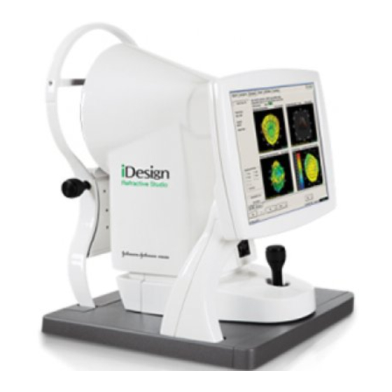
iDesign Advanced WaveScan Studio System 1. Keyboard 4. USB Port 2. Table Control 5. Main Power Switch 3. Joystick / Button 6. Printer Figure 5.1 – iDesign System iDesign System Model Eyes Two Model Eyes with a calibrated fixed diopter value are shipped with the instrument. The Model Eyes are used to confirm the proper operation and calibration of the instrument by measuring sphere, cylinder, and keratometry values daily. -
Page 25: Glide Pad
iDesign Advanced WaveScan Studio System Glide Pad To enter data with the Glide Pad, move the cursor to the appropriate box, left click once to activate the field, and type in the desired text. Left click once to use tabs or buttons. A single tap on the Glide Pad equals a left click. -
Page 26: Principle Of Operation
iDesign Advanced WaveScan Studio System Principle of Operation The Hartmann-Shack sensor measures the refractive error of the eye. The instrument evaluates the deflection of rays emanating from a small beam of light projected onto the retina. The system incorporates a fogged fixation target to control the eye’s natural accommodation during the imaging. The corneal topography function uses a spot reflection-based approach where the iDesign System analyzes the spot pattern image on the cornea and computes the corneal elevation and corneal power. -
Page 27: Enabling/Disabling The Hipaa Privacy Screen
iDesign Advanced WaveScan Studio System 1. Main Power Switch Figure 5.2 – Main Power Switch 1. System Power Switch Figure 5.3 – System Power Switch Enabling/Disabling the HIPAA Privacy Screen A privacy screen is displayed (Figure 5.4), which hides the current iDesign System screen after ten minutes of inactivity (U.S. -
Page 28: Software Description
iDesign Advanced WaveScan Studio System NOTE: In addition, if password protection is enabled, when the application starts, the privacy screen is displayed in locked mode. Figure 5.4 – System Privacy Screen Software Description The iDesign System software is designed specifically for the iDesign System. The software includes a Help System. -
Page 29: Software Navigation
iDesign Advanced WaveScan Studio System Software Navigation The tab-driven iDesign System software displays the available actions on tabs and buttons. Software Help System A green arrow guides the user to the next appropriate action. A red arrow prompts the user for missing or invalid required information. -
Page 30: Software Hierarchy
iDesign Advanced WaveScan Studio System Software Hierarchy The iDesign System software program features six main tabs at the top of the screen: About, Acquire, Review, Treat, Utilities, and System. • Select the About tab to display the software name and version number. •... -
Page 31: Acquire Tab
iDesign Advanced WaveScan Studio System Acquire Tab The Acquire tab (Figure 5.6) allows you to enter and modify patient information and to acquire patient measurements (see Section 8 – Acquiring Measurements) Figure 5.6 – Acquire Tab Screen System Functional Overview 0110-0624 Rev. -
Page 32: Review Tab
iDesign Advanced WaveScan Studio System Review Tab The Review tab (Figure 5.7) allows you to review existing patient data by selecting a patient and an exam, and then selecting the desired views from the Add Custom View menu. Refer to Section 9 – Reviewing Patient Data for a full description of its Review functionality. -
Page 33: Treat Tab
iDesign Advanced WaveScan Studio System Treat Tab The Treat tab (Figure 5.8) is used to design a Advanced CustomVue patient treatment based on a selected iDesign System exam. Refer to Section 10 – Designing Advanced CustomVue Treatments for a detailed description. Figure 5.8 –... -
Page 34: Utilities Tab
iDesign Advanced WaveScan Studio System Utilities Tab The Utilities tab (Figure 5.9) contains: • Clinic Settings • Treat Settings • Treat Adjustments • Database Maintenance - Sub-tabs are: Backup Database, Restore Database, Import Patient Data, Export Patient Data, Archive Patient Data, Merge Patient Records Delete Patients &... -
Page 35: System Tab
iDesign Advanced WaveScan Studio System System Tab The System tab (Figure 5.10) contains the Shutdown, System Settings, and System Maintenance sub- tabs used for shutting down the iDesign System software, setting the system date/time, setting printer preferences, setting mouse/glide pad user preferences, viewing system information, and performing software updates to the iDesign System software. -
Page 36: Using Usb Flash Drives
iDesign Advanced WaveScan Studio System Figure 5.11 – System, System Maintenance Sub-Tab Screen Using USB Flash Drives One USB port is located at the front of the iDesign System computer table, facing the user (see Figure 5.1). When connecting a USB flash drive to the iDesign System computer system for the first time, you may experience a lengthy delay (up to two minutes) for the system to recognize that the flash drive has been connected. -
Page 37: Removing A Usb Drive From The Idesign System Computer
iDesign Advanced WaveScan Studio System Removing a USB Drive from the iDesign System Computer To safely remove a USB flash drive from the iDesign System computer, click on the Eject USB Key (flash drive) button located at the bottom of the iDesign System screen. A pop-up screen appears with the following message: USB key successfully ejected. - Page 38 iDesign Advanced WaveScan Studio System Click on the Stop button. The Stop a Hardware device screen will appear. The USB Mass Storage Device is already selected on the screen. Click OK. 3. A screen will pop up with the message “The USB Mass Storage Device device can now be safely removed from the system”.
-
Page 39: Setting User Defaults
iDesign Advanced WaveScan Studio System Operator’s Manual Section 6 — Setting User Defaults Using the Utilities Options The Utilities tab contains the Clinic Settings, Treat Settings, Treat Adjustments, and Database Maintenance sub-tabs for setting personnel defaults, password options, refraction defaults, treatment types, treatment adjustment defaults, and for backing up and retrieving patient data from the database. - Page 40 iDesign Advanced WaveScan Studio System Operator’s Manual 1. From the Utilities tab, select the Clinic Settings sub-tab at the bottom of the screen. 2. Select the Enter/Edit Clinic Name box to enter the clinic name. The clinic name will be printed on all patient reports.
-
Page 41: Enabling/Disabling The Hipaa Privacy Screen
iDesign Advanced WaveScan Studio System Operator’s Manual Enabling/Disabling the HIPAA Privacy Screen A privacy screen is displayed (Figure 6.2), which hides the current iDesign System screen after ten minutes of no activity (U.S. HIPAA [Health Insurance Portability and Accountability Act]). If password protection is inactive, click OK to remove the privacy screen and return to the prior state of the application. -
Page 42: Setting/Modifying Default View Options
iDesign Advanced WaveScan Studio System Operator’s Manual Figure 6.3 – Password Entry Window To disable password protection, uncheck the HIPAA Password Enable check box. Enter the password for confirmation (Figure 6.4), and then click OK. Figure 6.4 – Verification for Disabling Password Protection Setting/Modifying Default View Options All available image types used in the iDesign System software have one or more viewing options. - Page 43 iDesign Advanced WaveScan Studio System Operator’s Manual Figure 6.5 – Utilities Tab, Clinic Settings Sub-Tab View Options can be set for the following maps and/or images: • Eye Image (Scotopic and Photopic Iris images with Pupilometry data) • Hartmann-Shack (HS) Image •...
-
Page 44: Setting/Modifying Default Treatment Settings
iDesign Advanced WaveScan Studio System Operator’s Manual Setting/Modifying Default Treatment Settings When you first modify a field in either the Treat Settings or Treat Adjustments sub-tab, a dialog box will appear (Figure 6.6) asking you to type “yes” in the text field and then click OK to allow the settings to be modified. - Page 45 iDesign Advanced WaveScan Studio System Operator’s Manual Figure 6.8 – Treat Settings Sub-Tab Set the following default surgical parameters. • Acuity Format: US (20/xx), Italy (xx/10), Metric (6/xx), or Decimal (x.xx). • Treatment Type: LASIK. • Lane Length: Enter the lane length that was used to perform the patient’s manifest refraction. Lane length will be used to adjust the manifest refraction to optical infinity for comparison with the wavefront refraction.
-
Page 46: Determining Refracting Lane Length
iDesign Advanced WaveScan Studio System Operator’s Manual • Treat/Patient tab options: • Exams needed for treatment: 1 or 3 or more. • Default Treat Calculation Mode: Express or Expert. • Select the default treatment card part number. • Enter LASIK Flap diameter and Flap Thickness dimension preferences. The LASIK flap diameter value can range from 8.0 mm to 10.0 mm and the LASIK flap thickness can range from 90 µm to 180 µm. -
Page 47: Lane Length Graphical Examples
iDesign Advanced WaveScan Studio System Operator’s Manual Lane Length Graphical Examples Mirrored System (Lane Length = a + b) Mirrored System (Lane Length = a + b) Projector to Screen (Lane Length = a) Monitor (Lane Length = a) Setting User Defaults 0110-0624 Rev. -
Page 48: Setting/Modifying Default Treatment Adjustments
iDesign Advanced WaveScan Studio System Operator’s Manual Setting/Modifying Default Treatment Adjustments Select the Treat Adjustments sub-tab (Figure 6.9) at bottom of screen to specify optional Nomogram and Physician Adjustments defaults on age-group basis. Fields on this sub-tab cannot be left blank; if no adjustment value is desired, enter a zero in that field. -
Page 49: Configuring The Promotional Printout Information Screen
iDesign Advanced WaveScan Studio System Operator’s Manual • Adjust to manifest: sphere will be automatically changed to the patient’s manifest sphere value entered on the PreOp sub-tab, within a range of ±0.75 D. If the sphere adjustment is more than ±0.75 D, it will be limited to ±0.75 D and the user is notified that the automatic adjustment exceeded the allowable limit. - Page 50 iDesign Advanced WaveScan Studio System Operator’s Manual Figure 6.11 – Clinic Settings, Promotional Printout Information Screen Setting User Defaults 0110-0624 Rev. D 0217 6-12...
-
Page 51: Managing Patient Data
iDesign Advanced WaveScan Studio System Operator’s Manual Section 7 — Managing Patient Data Specifications for the External USB Hard Drive • Must be a hard-drive not a USB stick • The Drive must be pre-formatted with NTFS Archive will not work unless the drive is formatted to NTFS •... -
Page 52: Backing Up And Restoring The Idesign System Database
iDesign Advanced WaveScan Studio System Operator’s Manual Figure 7.1 – Database Maintenance Sub-Tab Backing up and Restoring the iDesign System Database The iDesign System database can be backed up on the internal backup drive and restored by selecting the Database Maintenance sub-tab at the bottom of the screen and then selecting the Backup Database or the Restore Database button. -
Page 53: Importing Data From An Idesign System
Importing Data from an iDesign System WARNING! AMO recommends that you do not attempt to import and merge patient data captured on two separate iDesign System instruments until you have been trained to use this feature to avoid incorrect merging of patient records NOTE: The iDesign System treatment planning software is only compatible with exams performed with the iDesign System and does not support the import of patient data from the WaveScan System. - Page 54 iDesign Advanced WaveScan Studio System Operator’s Manual Figure 7.2 – File Open Window 3. Select the appropriate patient database and click Open. The Database Import screen will appear (Figure 7.3). Patient data can be imported by selecting individual patient files and exams using Manual Tagging or the entire patient database can be imported using Batch Tagging (selecting all patients).
- Page 55 iDesign Advanced WaveScan Studio System Operator’s Manual Figure 7.3 – Database Import Screen 4. To manually tag a patient file for import, select the patient list entry by clicking on it. Select as many patient names as desired. A check will appear in the box next to the last name and a list of exams for that patient will appear in the Exam Date box.
-
Page 56: Exporting Patient Data
iDesign Advanced WaveScan Studio System Operator’s Manual For example, two different treatments can exist for the same eye, one in the source, and one in the destination database. The radio buttons allow the user to specify the desired outcome: • leave records on destination machine as is •... - Page 57 iDesign Advanced WaveScan Studio System Operator’s Manual 1. Export Anonymously check box Figure 7.4 – Database Export Screen To export patient data, perform the following steps: 1. Select the Export Patient Data button on the Utilities screen. The Database Export screen will appear (Figure 7.4).
- Page 58 iDesign Advanced WaveScan Studio System Operator’s Manual 3. Tag the individual exams for each patient to be exported by clicking on the exams in the Exam Date box. If no exams are tagged, then the patient record without any exam records will be exported. 4.
-
Page 59: Archiving Patient Data
iDesign Advanced WaveScan Studio System Operator’s Manual Archiving Patient Data IMPORTANT: Before archiving the database, ensure the amount of free disk space on the external USB hard drive is sufficient for the required amount of disk space needed to archive the database. For example, a database containing 1000 patients with 10 exams per patient (10,000 total exams) will require approximately 11 gigabytes of disk space to archive. -
Page 60: Restoring The Archive
iDesign Advanced WaveScan Studio System Operator’s Manual 3. Click the Browse button to locate the desired directory on the USB device, and then click the Archive button to begin the archive process. If you have archived to an external USB drive before, the system will default to the USB drive. -
Page 61: Merging Patient Records
iDesign Advanced WaveScan Studio System Operator’s Manual Merging Patient Records Click on the Merge Patient Records button on the Database Maintenance sub-tab under the Utilities tab to view the Patient Merge screen (Figure 7.7). Merging patient records is useful when two or more separate patient records are accidentally created for the same patient. - Page 62 iDesign Advanced WaveScan Studio System Operator’s Manual Select the Delete Patients and Exams button. The Patient/Exam Delete screen will appear (Figure 7.8). Patient data and exam records can be deleted by selecting individual patients and exams (Manual Tagging) or the entire patient database can be deleted (Batch Tagging) (Figure 7.9). Exams taken within the last 12 months cannot be deleted by Batch Tagging.
- Page 63 iDesign Advanced WaveScan Studio System Operator’s Manual Figure 7.9 – Delete Batch Tagging Screen Managing Patient Data 0110-0624 Rev. D 0217 7-13...
-
Page 64: Acquiring Measurements
iDesign Advanced WaveScan Studio System Operator’s Manual Section 8 — Acquiring Measurements WARNING! Patients must discontinue the use of soft contact lenses at least two weeks prior to examination and treatment. Hard (PMMA) or RGP lenses must be discontinued at least three weeks prior to examination and treatment, with stable keratometry and refraction. - Page 65 iDesign Advanced WaveScan Studio System Operator’s Manual Figure 8.1 – Acquire Tab, Patient Sub-Tab Screen Acquiring Measurements 0110-0624 Rev. D 0217...
-
Page 66: Adding A New Patient's Data
iDesign Advanced WaveScan Studio System Operator’s Manual Adding a New Patient’s Data Follow the instructions below to enter new patient data. 1. Select the New Patient button below the list of patient records. The Add Patient window launches (Figure 8.2). Figure 8.2 –... - Page 67 iDesign Advanced WaveScan Studio System Operator’s Manual • Enter additional information (optional). Information entered on the Notes tab displays in the Patient Notes box on the Patient screen. 3. Click OK to save the data. The entered patient is now selected in the Patient window. 4.
-
Page 68: Editing Existing Patient Data
iDesign Advanced WaveScan Studio System Operator’s Manual Editing Existing Patient Data Follow the instructions below to find, select, and edit existing patient records. 1. On the Patient screen under Patient Search, type the initial letters of the patient’s last name, first name, and/or ID number in the boxes provided (Figure 8.5). - Page 69 iDesign Advanced WaveScan Studio System Operator’s Manual Figure 8.6 – Patient Selected After Search 3. To edit the patient information, select the Edit Patient button. The Edit Patient window appears (Figure 8.7). Figure 8.7 – Edit Patient, Demographics Window Acquiring Measurements 0110-0624 Rev.
-
Page 70: Preparing The Examination Room
iDesign Advanced WaveScan Studio System Operator’s Manual 4. Modify the patient’s Demographics and Notes information. 5. Click OK to save the data. The edited patient is selected in the Patient window. Preparing the Examination Room The examination room lighting must be dim to allow the patient’s eyes to dilate naturally. It may be difficult to acquire the measurement if the examination room lighting is too bright. -
Page 71: Positioning The Patient For Examination
iDesign Advanced WaveScan Studio System Operator’s Manual 4. Give the following instructions to the patient: • Open eyes widely when requested and do not blink during the measurement. • Keep mouth closed and head motionless during the measurement. • Relax and blink between measurements to keep eyes moist. •... - Page 72 iDesign Advanced WaveScan Studio System Operator’s Manual To position the patient properly (Figure 8.9), perform the following adjustments: 1. Adjust the chair height so that the patient’s back is straight, feet are flat on the floor, with thighs supported by the front of the seat cushion. 2.
-
Page 73: Eye Alignment And Focus
iDesign Advanced WaveScan Studio System Operator’s Manual Eye Alignment and Focus The iDesign System provides for a focusing adjustment (Z axis) moving toward or away from the patient; a horizontal positioning adjustment (X axis), and a vertical positioning adjustment (Y axis). All three axes are adjusted using the Joystick, although major adjustments for X and Z axes can be made by moving the measurement head. - Page 74 iDesign Advanced WaveScan Studio System Operator’s Manual To adjust vertical position (Y axis) of the measurement head: Rotate the Joystick clockwise to move the measurement head up, or counterclockwise to move the measurement head down. To focus and center for measurement: Move the Joystick forward toward the patient or backward away from the patient so the fiducial marks are in clear focus.
-
Page 75: Automatic Refraction (Precompensation)
iDesign Advanced WaveScan Studio System Operator’s Manual WARNING! Observe caution when moving the Joystick forward toward the patient. Avoid contact of the aperture and/or measurement head with the patient’s head. Automatic Refraction (Precompensation) After obtaining proper focus and alignment, select the automatic refraction function by pressing the Joystick button or using a left mouse click on the Auto Refract button.The automatic refraction function precompensates the instrument to the patient’s spherical equivalent and brings the fixation target into clear focus for the patient. -
Page 76: Capture
iDesign Advanced WaveScan Studio System Operator’s Manual Capture 1. After the automatic refraction cycle is complete, the instrument is ready to capture the measurement. Instruct the patient to blink once or twice, and then instruct them to keep their eyes open widely and gaze beyond the fixation target. - Page 77 iDesign Advanced WaveScan Studio System Operator’s Manual 1. Pupil outline (cyan circle) 2. Corneal vertex marker (yellow diamond) 3. Center of outer iris boundary (magenta cross) and pupil center (cyan cross) 4. Outer iris boundary (magenta circle) Figure 8.13 – Eye Image that Passes Analysis •...
-
Page 78: Screening Mode Procedure
iDesign Advanced WaveScan Studio System Operator’s Manual Screening Mode Procedure Use the Screening mode to capture one exam for pre-operative and/or post-operative evaluation. As you progress through the acquisition process, a check mark is displayed next to each completed tab and you are automatically moved to the next screen. - Page 79 iDesign Advanced WaveScan Studio System Operator’s Manual 5. Click the Next button or the OD Capture sub-tab at the bottom of the screen to begin the examination. Green arrows guide you to the next appropriate step. Helpful hints at the bottom left of the screen helps lead you through the acquisition process.
- Page 80 iDesign Advanced WaveScan Studio System Operator’s Manual Figure 8.16 – Acquire Tab, OD Image Review Sub-tab, Hartmann-Shack and Corneal Topography Image 7. Select the Next button, the OD Maps sub-tab, or the Joystick button to move to the OD Maps screen.
- Page 81 iDesign Advanced WaveScan Studio System Operator’s Manual Figure 8.17 – Acquire Tab, OD Maps Sub-tab, Viewing Maps of Higher Order Aberrations and Corneal Topography Elevation and Axial Power 8. To display the wavefront-derived values at a different diameter than specified in the defaults, select the desired diameter from the Pupil Size for Rx Calc panel.
-
Page 82: Treatment Mode Procedure
iDesign Advanced WaveScan Studio System Operator’s Manual Figure 8.18 – Map Report NOTE: You must continue to the OS Capture screen to save and print the OD Maps screen. If you do not need to capture OS, press the Skip OS button after you proceed to the OS Capture screen. The Capture OS, Review OS, and View OS maps procedure is identical to the procedure for OD. - Page 83 iDesign Advanced WaveScan Studio System Operator’s Manual 1. Select the Acquire tab when you are ready to begin the examination. The Patient screen will be displayed (Figure 8.19). 1. Select Treatment Mode Figure 8.19 – Start Acquisition in Treatment Mode 2.
- Page 84 iDesign Advanced WaveScan Studio System Operator’s Manual 6. Enter the keratometry values within the following ranges (required fields): • K1 = 30.0 D to 70.0 D • K2 = 30.0 D to 70.0 D • K2 Axis = 0° to 180° 7.
- Page 85 iDesign Advanced WaveScan Studio System Operator’s Manual Figure 8.20 – Pre-Op Screen for Acquisition in Treatment Mode 12. Select Next, or OD Capture at the bottom of the screen (Figure 8.20). You can also press the button on the Joystick to move through the acquisition procedure. Acquiring Measurements 0110-0624 Rev.
- Page 86 iDesign Advanced WaveScan Studio System Operator’s Manual Figure 8.21 – Capture Screen in Treatment Mode NOTE: The Save check box on the Exam List is checked when exams are captured successfully. With or without Pre-op information entered, exams may automatically be selected for treatment, but Pre-op information must be entered before calculating a treatment.
- Page 87 iDesign Advanced WaveScan Studio System Operator’s Manual Figure 8.22 – Capture Screen in Treatment Mode after successfully capturing three exams, with one exam selected for treatment 13. Continue capturing measurements until one of the exams is selected for treatment. Figure 8.22 shows an example of an exam selected for treatment.
- Page 88 iDesign Advanced WaveScan Studio System Operator’s Manual 16. Select Next after the exam is selected for Advanced CustomVue treatment. • If Pre-Op information has not been entered, the Acquire/Patient sub tab will appear. • If Pre-Op information has already been entered, the Treat/Pre-Op sub tab will appear, where you can modify Pre-Op information, if necessary, and then proceed with the treatment calculation.
-
Page 89: Troubleshooting During Treatment Mode Acquisition
iDesign Advanced WaveScan Studio System Operator’s Manual Troubleshooting during Treatment Mode Acquisition The Acquire tab capture screen in the Treatment mode features a Hints area, showing possible reasons for exam failure and suggestions for improving the capture rate. See next section for a list of messages and suggestions to improve the capture rate. -
Page 90: Suggestions To Improve Capture Rate
Advanced WaveScan Studio System Operator’s Manual Suggestions to Improve Capture Rate NOTE: If the suggested Hints do not resolve the issue, contact your AMO Customer Service representative. Message/ Image Message/Hints Image Hints HS area too small, Too few Eyelids too close to or HS points, Pupil detection overlapping pupil. - Page 91 Dim, unusable HS pattern. If Pupil diameter too small. this occurs often with different Hint: Pupil diameter too small patients, contact AMO for treatment calculation. Dim customer service. room lights, wait, and then attempt to capture eye again. Eye irregularities may result in...
- Page 92 If tear film breakup or bad focus. consistent across patients, call Ask the patient to blink and AMO Customer Service. open wide prior to capture. Hint: Not enough Helmholtz Hint: Unable to associate the spots were detected. This may cone spots with the eye image.
-
Page 93: Reviewing Patient Data
iDesign Advanced WaveScan Studio System Operator’s Manual Section 9 — Reviewing Patient Data Data Selection To access stored measurement data, click on the Review tab and perform the following steps. 1. Select the patient and exam you wish to review. 2. -
Page 94: Review Tab Functions
iDesign Advanced WaveScan Studio System Operator’s Manual Figure 9.1 – Review Tab, Patient Screen, Add Custom View Pop-up Menu Review Tab Functions To remove views from the Review tab, follow the steps below. Select Remove to remove the current view from the screen or click the arrow next to Remove to select Remove Current View or Remove All Views. -
Page 95: Printing Data
iDesign Advanced WaveScan Studio System Operator’s Manual The icons shown above are displayed right-justified in an exam’s list-entry cell to indicate the information listed below. Three icons are used to indicate the status of various parts of the exam on screens and printouts. - Page 96 iDesign Advanced WaveScan Studio System Operator’s Manual • Print Current View: prints the report as described above. • Print Preview Current View: displays a print preview screen of the report. • Print All Views: prints a report of all currently open views. NOTE: The printouts of the Hartmann-Shack and PSF views are reversed (black images on white background) to conserve printer ink and enhance the visibility of the images.
-
Page 97: Overriding Default Viewing Options For Data And Maps
iDesign Advanced WaveScan Studio System Operator’s Manual Overriding Default Viewing Options for Data and Maps Default view options are set under the Utilities/Clinic Settings tab (refer to Section 6 – Setting User Defaults). These view option defaults can be overridden for any view by selecting the View Options button inside the view and will affect only the currently selected view until you exit the iDesign System software application. -
Page 98: Wavefront Error And Refractive Correction Maps Difference
iDesign Advanced WaveScan Studio System Operator’s Manual Wavefront Error and Refractive Correction Maps Difference Unless otherwise indicated, the following options apply to both the Wavefront Error maps (WFR) and the Refractive Correction maps (Rx): • Show mm Grid • Show Angle Scale (2D Maps Only) •... -
Page 99: Corneal Topography Maps
iDesign Advanced WaveScan Studio System Operator’s Manual Corneal Topography Maps The user is able to set the following options independently for the CT Axial Power maps and the CT Best-Fit Sphere Elevation maps: • Show mm Grid • Show Angle Scale Scaling Options: •... -
Page 100: Components Of The 3D Plots
iDesign Advanced WaveScan Studio System Operator’s Manual Components of the 3D Plots When 3D images are displayed (Figure 9.2), you can rotate the image with a left-button mouse click- and-drag operation while the mouse cursor is inside the plot. Releasing the mouse button stops the rotation and leaves the plot in its current position. -
Page 101: Custom View
iDesign Advanced WaveScan Studio System Operator’s Manual • For Refractive Correction and Difference Maps, the range is displayed as the actual minimum and maximum value in the z direction, such as -xx.xx to +xx.xx diopters. • For PSF images, the range in minutes of arc are covered by the x and y axes, such as -xx.x to +xx.x minutes of arc. -
Page 102: Custom View Options
iDesign Advanced WaveScan Studio System Operator’s Manual Custom View Options By clicking on the View Options button, you can set your preference for maps and PSF images to be displayed as either two-or three-dimensional. Check the 3D box to activate the 3D display. The same map can be displayed as both 2D and 3D in the same Custom View. -
Page 103: Wavefront Error Maps
iDesign Advanced WaveScan Studio System Operator’s Manual Wavefront Error Maps Wavefront Error Maps (Figure 9.5) are a pair of maps for one or two exams. One or two exams can be displayed simultaneously with two maps per exam (up to four maps total) on one screen. A circular overlay depicting the user-selected calculation pupil is visible only if the user-selected pupil diameter is less than Max Pupil. -
Page 104: Wavefront Error Map View Options
iDesign Advanced WaveScan Studio System Operator’s Manual Wavefront Error Map View Options Viewing options for the selected map can be set by clicking on the View Options button displayed on the screen (Figure 9.6). • Show mm Grid: for 2-D maps, this enables the graphic overlay of an orthogonal millimeter grid over the map. -
Page 105: Wavefront Error Map Ansi Compliance
iDesign Advanced WaveScan Studio System Operator’s Manual The system will generate the following data: • Measured refraction (sphere, cylinder, axis, vertex) at the selected pupil diameter (2, 3, 4, 5, 6, 7, 8 mm or Max Pupil). • Pupil diameter used to calculate the above wavefront-based refraction. •... -
Page 106: Exam Selection Order
iDesign Advanced WaveScan Studio System Operator’s Manual The Wavefront Error Difference Maps (Figure 9.7) allow the clinician to compare the point-by-point differences between two exams of the same eye, Ex 1 and Ex 2. These difference maps are obtained by point-by-point subtraction of the corresponding individual maps. -
Page 107: Wavefront Error/Refractive Correction Difference Map View Options
iDesign Advanced WaveScan Studio System Operator’s Manual Wavefront Error/Refractive Correction Difference Map View Options Viewing options for the selected map can be set by clicking on the View Options button displayed on the screen (Figure 9.8). • Show mm Grid: for 2-D maps, this enables the graphic overlay of an orthogonal millimeter grid over the map. -
Page 108: Reading Wavefront Error Maps
iDesign Advanced WaveScan Studio System Operator’s Manual Reading Wavefront Error Maps The iDesign Advanced WaveScan Studio System displays the sphero-cylindrical refractive errors and high order wavefront aberrations as the optical path difference (OPD) between the measured outgoing wavefront and the ideal plane wavefront. Regions of the pupil with positive OPD are in front of the ideal plane and are shown in red. -
Page 109: Selecting Pupil Diameter For Calculating Wavefront-Based Refractions
iDesign Advanced WaveScan Studio System Operator’s Manual Selecting Pupil Diameter for Calculating Wavefront-based Refractions Changing the pupil diameter (Pupil Size for Rx Calc) on the maps screen will recalculate the sphere, cylinder, Hi Order %, RMS Error, and Eff Blur of the wavefront-based refraction to simulate changes in pupil size. -
Page 110: Point Spread Functions Image View Options
iDesign Advanced WaveScan Studio System Operator’s Manual Figure 9.9 – Point Spread Function Image View in 3D for Both Eyes Point Spread Functions Image View Options Viewing options (Figure 9.10) for the selected image can be set by clicking on the View Options button displayed on the screen. - Page 111 iDesign Advanced WaveScan Studio System Operator’s Manual Figure 9.10 – View Options, Point Spread Function Tab Reviewing Patient Data 0110-0624 Rev. D 0217 9-19...
-
Page 112: Zernike Coefficients Table
iDesign Advanced WaveScan Studio System Operator’s Manual Zernike Coefficients Table Zernike coefficients from the second to the sixth order are displayed in the Zernike Coefficients Table (Figure 9.11). The coefficients are calculated at the currently selected pupil size, and that value is displayed as part of the table header. -
Page 113: Zernike Coefficients Difference Table
iDesign Advanced WaveScan Studio System Operator’s Manual Zernike Coefficients Difference Table The difference in Zernike coefficients between two exams of the same eye are displayed in the Zernike Coefficients Difference Table (Figure 9.12). This view allows the clinician to compare the normalized Zernike coefficients of a pair of exams (Ex 1 and Ex 2) of the same eye by displaying the coefficients for each exam, as well as their coefficient differences obtained by subtraction (Ex 2 –... -
Page 114: Zernike Coefficients Difference Table View Options
iDesign Advanced WaveScan Studio System Operator’s Manual Zernike Coefficients Difference Table View Options Viewing options (Figure 9.13) for the selected table can be set by clicking on the View Options button displayed on the screen. You can select up to two exams for any patient, and open up a view containing the value of the normalized Zernike coefficients for the exam(s), dimensioned in microns (µm). -
Page 115: Corneal Topography Elevation Map
iDesign Advanced WaveScan Studio System Operator’s Manual Corneal Topography Elevation Map The Corneal Topography BFS (best-fit sphere) Elevation map can be displayed as part of a Custom View or a Corneal Topography Difference Maps view (Figure 9.16). This map consists of the following components: •... - Page 116 iDesign Advanced WaveScan Studio System Operator’s Manual Figure 9.14 – View Options, CT Maps Tab, Elevation Map Options Two scaling options are available for the CT Elevation Sphere-Difference Maps: • Auto-Scale: the step size of the color legend is chosen automatically to maximize the number of colors used in the map.
-
Page 117: Corneal Topography Axial Power Map
iDesign Advanced WaveScan Studio System Operator’s Manual Corneal Topography Axial Power Map The Corneal Topography Axial Power map can be displayed as part of a Custom View or a Corneal Topography Difference Maps view (Figure 9.16). This map consists of the following components: •... - Page 118 iDesign Advanced WaveScan Studio System Operator’s Manual Two scaling options are available for the CT Axial Power map: • Auto-Scale (non ANSI standard): both step size and central interval of the color legend are chosen automatically to maximize the number of colors used in the map. The map’s legend has 21 colors varying gradually from blue to red, with green for the center interval.
-
Page 119: Corneal Topography Difference Maps
iDesign Advanced WaveScan Studio System Operator’s Manual Corneal Topography Difference Maps The Corneal Topography Difference Maps (Figure 9.16) allow the clinician to compare a pair of Corneal Topography Axial Power exams or a pair of Elevation Difference exams by displaying the difference maps between an exam [Ex 1] taken at a specified time [t1], and an exam [Ex 2] taken of the same eye at a later time [t2]. -
Page 120: Corneal Topography Difference Maps View Options
iDesign Advanced WaveScan Studio System Operator’s Manual Corneal Topography Difference Maps View Options The following view options are available for this view (Figure 9.17): • Show Grid: when checked, this enables the graphic overlay of an orthogonal millimeter grid over the map. -
Page 121: Eye Image View
iDesign Advanced WaveScan Studio System Operator’s Manual Eye Image View Two types of Eye Image views can be displayed: • The Ambient Light Eye Image: displays ambient-light eye image. • The Photopic Eye Image: displays the high-illumination eye image. The two views are distinguished by the view title, and an icon overlay in the lower left corner of the image: A Moon symbol for the Ambient Light Eye Image. - Page 122 iDesign Advanced WaveScan Studio System Operator’s Manual In addition, the ambient light eye image will display the usability status for Iris Registration in the lower right corner of the image. Eye Image Usability Status for Iris Registration Unusable for treatment. Usable for treatment, but with a lower probability of successfully using Iris Registration on the STAR S4 IR Excimer Laser System.
-
Page 123: Eye Image View Options
iDesign Advanced WaveScan Studio System Operator’s Manual Eye Image View Options Viewing options (Figure 9.19) for the optional overlays for both the Ambient Light and Photopic Eye Image views can be set by clicking on the View Options button displayed on the screen. The check boxes control the marker visibility everywhere that the Eye Image can be displayed. -
Page 124: Hartmann-Shack (Hs) Images
iDesign Advanced WaveScan Studio System Operator’s Manual Hartmann-Shack (HS) Images The Hartmann-Shack view (Figure 9.20) displays the Hartmann-Shack image associated with an exam in its proper orientation. Each image has a color-coded “W” icon in its lower right corner. The color of the icon indicates the status of the image. -
Page 125: Hartmann-Shack Image View Options
iDesign Advanced WaveScan Studio System Operator’s Manual Figure 9.20 – Hartmann-Shack Review Screen Hartmann-Shack Image View Options Wavefront Circular Outline: When the Show check box is checked, the wavefront circular outline is displayed with a user-selectable color (default is green). Reviewing Patient Data 0110-0624 Rev. -
Page 126: Corneal Topography Image
iDesign Advanced WaveScan Studio System Operator’s Manual Corneal Topography Image A Corneal Topography image (Figure 9.21) is available in its own view. An icon is displayed in the lower right corner. When the icon is green, the corneal topography image analysis has succeeded. On top of the image, the standard 3-mm zone Keratometry values derived from the image analysis are displayed. -
Page 127: Designing Advanced Customvue Treatments
iDesign Advanced WaveScan Studio System Operator’s Manual Section 10 — Designing Advanced CustomVue Treatments Introduction Advanced CustomVue treatments are designed from the Treat tab either by selecting the Treat tab or moving to the Treat tab automatically from the Acquire tab using the Treatment Mode feature on the Acquire tab. -
Page 128: Selecting The Appropriate Idesign System Exam For Advanced Customvue Treatments
iDesign Advanced WaveScan Studio System Operator’s Manual Selecting the Appropriate iDesign System Exam for Advanced CustomVue Treatments The automated exam selection algorithm is designed such that the user has the ability to keep acquiring (or selecting) exams in order to improve the quality of the exam selected for treatment. After an exam has been selected, more exams can be captured (or selected by the user), and the selection algorithm is run again to select an exam out of the newly augmented pool of valid exams. - Page 129 iDesign Advanced WaveScan Studio System Operator’s Manual The pre-determined tolerance range is listed below: • Sphere Equivalent (SE): 0.625 > (MRSE – wavefront SE) > -0.625 D. • Cylinder: Delta Cylinder <=+/- 0.5 D. • Cylinder Axis: Both cylinders must have a magnitude of at least 0.5 D, or else the Axis difference test is ignored;...
-
Page 130: Selecting Idesign System Exams For Treatment
iDesign Advanced WaveScan Studio System Operator’s Manual Selecting iDesign System Exams for Treatment The user may select several exams and have the iDesign System software select the optimal exam to use for treatment by following these steps: 1. Click on the Treat tab. The Patient sub-tab will appear. 2. - Page 131 iDesign Advanced WaveScan Studio System Operator’s Manual Figure 10.1 – Treat Tab, Patient Sub-Tab Showing One Exam Needed Designing Advanced CustomVue Treatments 0110-0624 Rev. D 0217 10-5...
- Page 132 iDesign Advanced WaveScan Studio System Operator’s Manual 3. A single exam can also be chosen for treatment (if the single exam for treatment is the default setting) on the Review tab. Check the Use to Treat box in the upper right corner of the selected Wavefront Error/Refractive Correction Map sub-tab or the Custom View sub-tab (refer to Figure 10.2 and Figure 10.3).
-
Page 133: Preop Sub-Tab Options
iDesign Advanced WaveScan Studio System Operator’s Manual PreOp Sub-Tab Options The PreOp sub-tab (Figure 10.4) is used to enter preoperative information for one or both eyes. You can enter preoperative information after capturing exams, but all information must be entered before selecting an exam to be used for treatment. -
Page 134: Treatment Adjustments For The Idesign System
iDesign Advanced WaveScan Studio System Operator’s Manual Treatment Adjustments for the iDesign System Two types of adjustments are available for designing Advanced CustomVue treatments: the Physician Adjustment and the Percentage Nomogram Adjustment. The Percentage Nomogram Adjustment allows the surgeon to make a pre-defined percentage adjustment to the entire Advanced CustomVue-defined ablation. -
Page 135: Using Expert And Express Modes
iDesign Advanced WaveScan Studio System Operator’s Manual Using Expert and Express Modes Expert Mode (Figure 10.6) displays a Design and Calc sub-tab for each eye (OD/OS). In this mode, the design and calculation of a treatment is performed for one eye at a time. Express Mode (Figure 10.7) combines the individual Design and Calc sub-tabs used in Expert Mode into one Treat sub-tab that displays both eyes (OD/OS) on one page. - Page 136 iDesign Advanced WaveScan Studio System Operator’s Manual Figure 10.6 – Designing an Advanced CustomVue Treatment in Expert Mode Designing Advanced CustomVue Treatments 0110-0624 Rev. D 0217 10-10...
- Page 137 iDesign Advanced WaveScan Studio System Operator’s Manual Figure 10.7 – Treat/Patient Screen with Express Mode Selected Designing Advanced CustomVue Treatments 0110-0624 Rev. D 0217 10-11...
-
Page 138: Entering Treatment Design Parameters
iDesign Advanced WaveScan Studio System Operator’s Manual Figure 10.8 – Designing an Advanced CustomVue Treatment in Express Mode Entering Treatment Design Parameters The Advanced CustomVue treatment parameters from the selected wavefront measurement will be shown on the OD/OS Design screen (Figure 10.9). The OD/OS Design screen shows interactive displays of the following: •... - Page 139 iDesign Advanced WaveScan Studio System Operator’s Manual 1. Zoom button Figure 10.9 – Treat/OD Design Sub-Tab Screen in Expert Mode Designing Advanced CustomVue Treatments 0110-0624 Rev. D 0217 10-13...
- Page 140 iDesign Advanced WaveScan Studio System Operator’s Manual Figure 10.10 – Treat/Treat Sub-Tab Screen in Express Mode The Eye Image (Figure 10.11) has three main states displayed in the lower right corner of the image: Unusable for treatment. Usable for treatment, but with a lower probability of successfully using Iris Registration on the STAR S4 IR Excimer Laser System.
- Page 141 iDesign Advanced WaveScan Studio System Operator’s Manual Figure 10.11 – Zoom Feature Popup Screen for Eye Image Enter or confirm the following surgical treatment parameters: • Enter Physician Adjustments to the Advanced CustomVue treatment, if appropriate. The allowed Physician Adjustment limits for sphere are ±0.75 D. The Physician Adjustment is intended to be used as an endpoint adjustment to deviate from emmetropia.
- Page 142 iDesign Advanced WaveScan Studio System Operator’s Manual • Select the appropriate treatment card part number (Expert Mode only). Click on the down arrow to view a list of the available treatment card part numbers. • Surgical Parameters: • LASIK Flap Diameter (mm) Range 8 mm -10 mm •...
-
Page 143: Calculating The Advanced Customvue Treatment
iDesign Advanced WaveScan Studio System Operator’s Manual Calculating the Advanced CustomVue Treatment 1. Click on the OD/OS Calc sub-tab. 2. Click the Start button to begin the treatment calculation process. The Calculation Progress bar will indicate when the calculation is in progress, which may require several seconds to complete. When the treatment calculation is complete, the status box will display Calculation Complete. - Page 144 iDesign Advanced WaveScan Studio System Operator’s Manual Table 10.1 – Tolerance Range Between Manifest and Wavefront Refraction for Treatment Exam/Manifest Comparison Limits Minimum Maximum MRSE (adjusted to infinity) -wavefront SE -0.625 D 0.625 D MR Cylinder -wavefront cylinder -0.50D 0.50D Cylinder Axes, when both manifest and Decreases linearly from 15°...
- Page 145 iDesign Advanced WaveScan Studio System Operator’s Manual If the Advanced CustomVue calculation fails, review the treatment parameters entered on the OD/OS Design sub-tab. Table 10.3 displays the types of error messages that will appear in the Additional Information window on the OD/OS Calc sub-tab if the Advanced CustomVue calculation fails. After the Advanced CustomVue treatment has been successfully calculated, the following treatment statistics and information will be displayed on the OD/OS Calc sub-tab: •...
-
Page 146: Saving An Advanced Customvue Treatment
iDesign Advanced WaveScan Studio System Operator’s Manual Saving an Advanced CustomVue Treatment The calculated treatment plan must be saved to a USB flash drive to transfer the treatment plan to the STAR S4 IR Excimer Laser system. 1. Ensure a USB flash drive is inserted into the USB port of the iDesign System computer. Note: If a USB flash drive is present, the Express Mode automatically saves the treatment. - Page 147 iDesign Advanced WaveScan Studio System Operator’s Manual Surgical planning forms and surgical treatment plan reports are printed from the Treat tab in the Treatment Mode. If the Advanced CustomVue treatment has been calculated, clicking on the up arrow next to the Print button will display a pop-up menu with the following options: •...
-
Page 148: Surgical Planning Form
iDesign Advanced WaveScan Studio System Operator’s Manual Surgical Planning Form Figure 10.14 – Example of a Surgical Planning Form The surgical planning form (Figure 10.14), is used by the surgeon to specify the type of surgery, identify the iDesign System exam to be used as the basis for the surgery, and various other treatment parameters. If data have been entered into the iDesign System’s database using the Treat/PreOp sub-tab screen, then that data will be printed on the form. -
Page 149: Surgical Treatment Plan Report
iDesign Advanced WaveScan Studio System Operator’s Manual Surgical Treatment Plan Report Figure 10.15 – Example of a Surgical Treatment Plan Report The surgical treatment plan report (Figure 10.15) is intended to be printed after the Advanced CustomVue treatment calculations have been completed. It contains a summary of the planned surgical treatment, one eye per printed form, and all the available pre-operative and operative parameters. -
Page 150: Copying An Advanced Customvue Treatment To The Star S4 Ir Excimer Laser System
iDesign Advanced WaveScan Studio System Operator’s Manual Manifest and cycloplegic parameters are displayed for both current and infinite lane length. Spherical equivalent at infinity is displayed for both wavefront refraction (WF Rx) and manifest Rx. Note that the difference is only displayed if the vertex distance for WF Rx and manifest Rx is the same. The surgical treatment plan report can be printed from any of the Treat sub-tabs, provided the treatment has been calculated successfully. - Page 151 iDesign Advanced WaveScan Studio System Operator’s Manual 5. Click the first patient on the displayed list, which will highlight the selection with a dark blue background. 6. Hold down the SHIFT key, and then click on the last patient in the list. This action will result in every name in the list being selected and highlighted with a dark blue background.
-
Page 152: Self Test And Daily Verification
2. Select the System Maintenance sub-tab at the bottom of the screen. 3. Press Run Self-Test button to initiate the self test procedure. 4. Call AMO customer service if Fail appears in red next to a system component name. Self Test and Daily Verification 0110-0624 Rev. -
Page 153: Daily Verification Test
iDesign Advanced WaveScan Studio System Operator’s Manual Daily Verification Test Once per day, run the Daily Verification Test to verify that measurements of K1 and K2 keratometry, sphere and cylinder are within specification. The Daily Verification Test uses a Model Eye (depicted in figure 11-2) to ensure that the instrument is performing within the expected tolerance that were established in the factory. - Page 154 iDesign Advanced WaveScan Studio System Operator’s Manual Figure 11.3 – Model Eye Mounted on Chin Rest Figure 11.4 – Close-up of Model Eye on Chin Rest. 2. Press the System Power Switch on the front of the computer and allow the system to boot up. Self Test and Daily Verification 0110-0624 Rev.
- Page 155 iDesign Advanced WaveScan Studio System Operator’s Manual 1. System Power Switch Figure 11.5 – System Power Switch 3. Click the Acquire tab. Figure 11.6 – Main Screen 4. On the Acquire tab. Click on the TEST EYE patient or type in test under Patient Search – Last Name.
- Page 156 iDesign Advanced WaveScan Studio System Operator’s Manual 6. Select a Physician and Operator from the respective drop-down menus, and then click Next. Figure 11.7 – Acquire Tab, Patient Tab screen 7. Use the Joystick to align the pupil and focus the fiducials. NOTE: If the Purkinje reflections (four bright spots in the sample image below) DO NOT appear centered in the image’s pupil, check that the Model Eye is mounted correctly and is square to the optical head (see Figure 11.3).
- Page 157 iDesign Advanced WaveScan Studio System Operator’s Manual Topography and Illumination Topography and Illumination Purkinje Reflections with Fiducial Purkinje Reflections with Fiducial features (small + marks) features (small + marks) Figure 11.8 – Acquire Tab Screen, Purkinje Reflections 8. Click on Auto Refract. 9.
- Page 158 iDesign Advanced WaveScan Studio System Operator’s Manual Figure 11.9 – Acquire Tab, Hartmann-Shack and Corneal Topography Image NOTE: If a circle does not surround the Hartmann-Shack image’s spots or the green wavefront icon is missing from the image, select the OD Capture sub-tab and then repeat Steps 8 through 10. If the images still do not meet the criteria in this step, check the Model Eye’s position on the chin rest for tip and tilt.
- Page 159 iDesign Advanced WaveScan Studio System Operator’s Manual Figure 11.10 – OD Maps Screen 13. Verify the Pupil Size for RX Calc is set to 6 mm. 14. Compare the displayed iDesign System Values (example shown above) to the values printed on the Model Eye’s sticker.
- Page 160 17. Look into the aperture in the optical head. You should see the fixation target clearly and in focus. If the target does not appear, contact AMO Customer Service. 18. Click Skip OS or Skip OD. Self Test and Daily Verification 0110-0624 Rev.
-
Page 161: Troubleshooting
iDesign Advanced WaveScan Studio System Operator’s Manual Section 12 — Troubleshooting Warning Messages During Treatment Calculation The table below shows the appropriate corrective action to take for warning messages that may appear during treatment calculation. Warning Message Corrective Action Manifest SphEq. and WF SphEq differ by x.xxD Select Yes or No on the screen. -
Page 162: Warning Messages Displayed After Treatment Calculation
iDesign Advanced WaveScan Studio System Operator’s Manual Warning Messages Displayed After Treatment Calculation The table below displays the types of warning messages that may appear in the Additional Information window on the OD/OS Calc sub-tab after the Advanced CustomVue calculation has successfully completed. - Page 163 iDesign Advanced WaveScan Studio System Operator’s Manual Index Backup Database button barometric pressure Numerics operating Batch Patient & Exam Tagging screen 9-5, 9-17 2D images 7-4, 7-8, 7-12 Batch Tagging 9-5, 9-17 3D images 3D plots, components 8-13 Capture 11-6 Capture Button 9-13, 9-14, 9-16 aberrations...
- Page 164 iDesign Advanced WaveScan Studio System Operator’s Manual 7-11 11-6, 11-7, 11-8 Edit Target Patient check box Hartmann-Shack editing Hartmann-Shack Images 5-9, 6-1 8-16 operator name reviewing 5-9, 6-1 9-3, 9-13, 9-32 physician name Hartmann-Shack images 9-13, 9-16, 9-17 Effective Blur archiving electrical ratings printing...
- Page 165 iDesign Advanced WaveScan Studio System Operator’s Manual LASIK Flap diameter optical system lock mode optical zone dimensions 5-6, 6-3 10-19 privacy screen OS Calc sub-tab maintenance paper, setting print orientation 5-5, 6-3 system password protection 6-10, 8-20, 10-8, 10-17 5-6, 6-3 manifest refraction procedure differences between manifest and wavefront...
- Page 166 iDesign Advanced WaveScan Studio System Operator’s Manual 9-17 11-2 PSF images, definition of self test 11-1 PSF views, printing self test procedure 9-17 pupil diameter semi-meridians 9-17 selecting for calculations Show Data Loss Warning 6-2, 9-13, 9-16, 10-16 pupil size Show HS Tab 10-18, 12-1 10-12...
- Page 167 iDesign Advanced WaveScan Studio System Operator’s Manual Usability status 9-3, 9-13 Iris Registration USB external hard drive 5-16, 7-3, 7-8, 10-20 USB flash drives 5-16 USB Mass Storage Device 5-8, 11-1 Utilities tab Utilities/Clinic Settings 10-19 Variable Spot Scanning vertex distance 6-2, 9-5, 9-20 view options viewing data...
Need help?
Do you have a question about the iDesign G300 and is the answer not in the manual?
Questions and answers